![MIC values of aqueous plant extracts[68].](/storage/images/resized/iRgsDaHPJ4YdcvI4uP3aTzYnPsdGD8zl5tLU4OBY_xl.webp)
![Mean thickness of the E. faecalis biofilm[83].](/storage/images/resized/iEVDbfPMI3AveCYe4Yk706U7aq3AFfKeOncid0p3_xl.webp)
Keywords
Abstract
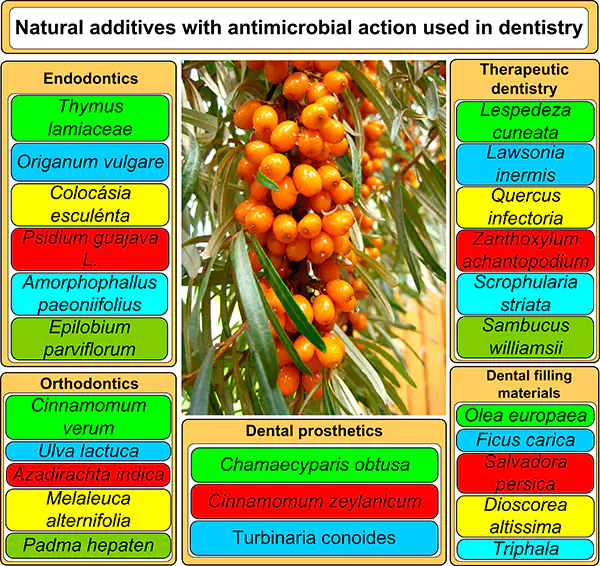
The existing additives for dental restorative materials used in therapeutic dentistry, dentures, orthodontic appliances and adhesives (for example, metal and metal oxide nanoparticles) and also additives to root canal irrigants used in endodontics and to mouthwashes can be toxic to humans, cause allergic reactions and accumulate in organs and tissues. Today, a relevant trend is development of dental materials that have an antimicrobial effect and are non-toxic to humans. A promising alternative to the above-mentioned additives are phyto-components, including plant and propolis extracts, since they are cheap and non-toxic. This review is devoted to natural antimicrobial additives to dental materials used in orthodontics, dentures, therapeutic dentistry and endodontics. The review makes recommendations regarding additional research required for practical implementation of the considered dental materials (filling materials, mouthwashes, orthodontic adhesives and orthopaedic dental products), examines the influence of antimicrobial additives on the physicochemical and physicomechanical properties of polymer dental materials, and outlines the advantages and disadvantages of natural additives compared to synthetic ones. The main challenges in this research area are the narrow range of microorganisms for which the antimicrobial effect was studied (which are mainly S. mutans, E. faecalis, C. albicans) and the predominance of in vitro studies over clinical studies. The review covers published data of the past five years.
The bibliography includes 123 references.
1. Introduction
Oral health has a great impact on person’s quality of life, health and aesthetic perception. Therefore, the development of dentistry is an important area of modern medicine. One of the most important problems faced by dentists is the detrimental effect of pathogenic and conditionally pathogenic microflora on the tissues of the oral cavity, both during normal human life and during treatment or aesthetic procedures. This influence can lead to various kinds of complications, for example, gingivitis, periodontitis, caries, denture stomatitis, peri-implantitis, etc.
Materials meant for dentistry should meet certain requirements, in particular, dental restorative materials should possess mechanical strength, low degree of shrinkage, chemical stability, low solubility; mouthwashes should have a refreshing smell and mild taste, no mechanical inclusions and no harmful effects on the human body; root canal irrigants should have high dissolving activity to canal dentin, low surface tension for delivery of the irrigant to the entire working length of the root canal, to hard-to-reach places and deep into the root canal and the lack of toxicity towards human dermal fibroblasts. In addition, dental materials must have a continuous antimicrobial effect against major oral microorganisms.
The main pathogenic and opportunistic microorganisms of the oral cavity include streptococci S. mutans, S. salivarius, S. sobrinus and S. sanguis, which cause caries, enterococcus E. faecalis, which is the main causative agent of root canal infections, gram-negative bacteria P. ginigivalis and grampositive bacteria A. naeslundii, involved in periodontal diseases and Candida fungi (C. albicans and C. glabrata species), which cause oral candidiasis, prosthetic stomatitis and peri-implantitis.
To combat opportunistic microflora in the oral cavity, many materials have been developed containing various antimicrobial additives: nanoparticles, synthetic chemicals and natural biologically active substances.
For example, for odontogenic infections that cause pathological changes in dental tissues, the antibiotic amoxicillin is effective [1]. A disadvantage of antibiotics, such as ampicillin, chloramphenicol, gentamicin, tetracycline and kanamycin, is the antibiotic resistance of opportunistic microorganisms of supragingival dental plaque [2].
Nanoparticles of metals, metal oxides and calcium fluoride [3], polymer nanoparticles based on chitosan, and poly (lactic-coglycolic acid) [4] are actively used as antimicrobial additives. This is due to the high surface area to volume ratio and high charge density, which allows for their effective interaction with bacterial cells [5]. Today, ‘nanodentistry’, which implies the use of nanomaterials in orthodontics, therapeutic dentistry and dental prosthetics, is an actively developing area [6]. Calcium fluoride nanoparticles in light-curing adhesives reduce the number of S. mutans bacteria after a month and decrease the enamel demineralization for a period of 6 months after bonding and after appliance removal (i.e. 6 months after placement until the appliance is removed) in orthodontic patients [7]. The addition of silver nanoparticles to light-curing acrylic resins endows them with a strong antibacterial effect against grampositive bacteria S. mutans [8]. Coating implants with silver nanoparticles not only protects them from the formation of S. mutans bacterium and Candida fungi biofilms and secondary infections, but also helps the implant integrate into bone tissue, increase the implant biocompatibility and make it more durable [9]. Gold nanoparticles reduce the extent of Candida albicans adhesion to poly(methyl methacrylate) (PMMA)-based dentures by 66% after 24 hours of observation [10], which is due to a smoother surface and higher electrostatic repulsion due to a lower z-potential compared to those of unmodified materials.
However, a drawback of nanoparticles is cytotoxicity against human fibroblast cells, DNA damage and the ability to accumulate in organs [11], which is due to their small size (less than 100 nm), large surface area, which increases their absorption by the cell and production of reactive oxygen species (ROS) [12]. Nanoparticles are prone to agglomeration, which leads to loss of antimicrobial activity. To solve the problem of agglomeration, nanoparticles must be stabilized with a polymer shell, which increases the cost of their synthesis [13].
Sodium perborate, sodium hypochlorite, glutaraldehyde, chlorine dioxide and chlorhexidine digluconate can be used as chemicals for denture disinfection. However, the disadvantage of these products is the risk of damage to the surface of prostheses based on acrylic resins, changes in the mechanical properties of the prosthesis and cytotoxicity. In particular, sodium perborate has a cytotoxic effect on human gingival fibroblasts [14]. In turn, treatment of dentin with chlorhexidine before bonding a dental restorative material to dentin, can reduce the shear bond strength between dentin and dental restorative material and impair the durability of the restoration [15]. Chlorhexidine digluconate mouthwash causes yellow-brown stains on the surface of dental restorations and thereby compromises their esthetics [16].
Recently, biologically active substances from natural sources have attracted increasing attention of researchers. Many of plant extracts are used in the food industry as food preservatives and antioxidants [17], [18], [19], [20], spicy and aromatic additives [21], in cooking [22], in cosmetology as part of skin care products [23], in traditional medicine as drugs with antibacterial, antidiabetic, antiinflammatory, antimalarial, antiviral, wound healing, hemostatic or immunomodulatory effects [24], [25], [26], [27]; in the biological synthesis (Biological synthesis of cellulose fibres is the isolation of native cellulose fibres from peels of A. vera.) of cellulose nanofibres [28], silver nanoparticles [29], copper and zinc oxides [30] as stabilizers and reducing agents. For example, an aqueous extract of the Mediterranean cypress (Cupressus sempervirens) leaves and aqueous extracts of hemp fibre [31] promotes the reduction of silver ions from an aqueous solution of silver nitrate to give silver nanoparticles [32]. An aqueous extract of Aloe vera leaves in contact with copper ions changes the colour of a copper(II) acetate monohydrate solution from blue to green [33], which indicates the formation of copper oxide nanoparticles from copper ions.
Owing to high content of phenolic compounds, in particular flavonoids, plant extracts of Scutellaria baicalensis, Yucca filamentosa, Dracocephalum moldavica L., Lamium galeobdolon L., Beilschmiedia alloiophylla, Beilschmiedia kunstleri and Vaccinium axillare Nakai, have an antioxidant effect, neutralize free radicals and reactive oxygen species, causing oxidative stress, which makes them promising for reducing the toxicity of cytostatic drugs [34], [35], treating cardiovascular, neurodegenerative, and gastrointestinal diseases [36], [37], [38], [39].
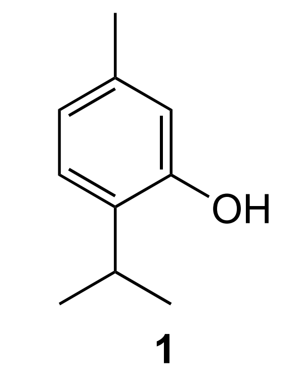
Natural biologically active substances are also used as antimicrobial additives in dentistry, as they contain secondary metabolites. Among the main mechanisms of antimicrobial action of secondary metabolites are oxidative stress, inhibition of cell wall synthesis, damage to the cell membrane, inhibition of nucleic acid and protein synthesis [40]. For example, the antibacterial and antifungal activities of thyme are due to the presence of thymol (2-isopropyl-5-methylphenol) (1), which is embedded in the lipid layer of the cell membrane of gramnegative bacteria (for example, E. coli) and interacts with the polar part of the membrane (polysaccharide with phosphate groups) via a hydroxyl group and with the hydrocarbon tail of the membrane (fatty acid residue) via the hydrophobic part (benzene ring and aliphatic side chain). This results in destabilization of the outer membrane lipid bilayer, leakage of the outer membrane, cell wall and cytoplasmic ions, change in the proton motive force of the bacterial cell and lysis of the contents (Figure 1, Figure 2) [41]. Also, thymol (Structure 1) reduces the concentration of ergosterol in the cell membrane of Candida fungi, increases the concentration of reactive oxygen species and causes oxidative stress, thus reducing the content of extracellular polymeric matrix (EPM), a capsular polysaccharide, and disrupting the integrity of the cell membrane (Figure 3) [41].
![[{"type":"paragraph","data":{"text":"Interaction of thymol with the cell wall of gram-negative bacteria."}}]](/storage/images/resized/fhj0vKcEOw3S2id3aoaLWgXP8053pax3s5Xfq872_xl.webp)
Natural additives, unlike antibiotics of the tricyclic glycopeptide group such as vancomycin, have antimicrobial effects against multi-drug resistant (Multi-drug resistance occurs when a pathogenic organism shows increased resistance to multiple antibiotics, making treatment difficult.) oral bacteria E. faecalis [42].
![[{"type":"paragraph","data":{"text":"Effect of thymol on gram-negative bacteria (I is leakage of outer membrane, II is change in the proton motive force of the bacterial cell, III is cell lysis)."}}]](/storage/images/resized/KyyT7lQUoob8axP6uLTcX5V0g8OKyvfyNpumylhu_xl.webp)
Compared to nanoparticles, additives of natural origin, including phyto-additives, are cheap, non-toxic to humans, and rarely cause allergic reactions [17].
![[{"type":"paragraph","data":{"text":"Effect of thymol on the fungal cell membrane."}}]](/storage/images/resized/ok0FvyQKcToAU4CyYAbyZDGLo7NYZhcus3Zc7nWy_xl.webp)
Unlike synthetic antimicrobial agents, natural additives are safer and do not cause significant side effects, such as changes in taste, colour of teeth, tongue or lips, peeling and inflammation of the oral mucosa [43].
The antimicrobial effect of plant extracts and dental materials based on them is studied using the diffusion method (well method), where Mueller Hinton agar acts as a nutrient medium. The effect of extracts on bacterial biofilms, in turn, is studied using a confocal laser microscope. Wells are applied to agar using a sterile drill, then Petri dishes are incubated with a nutrient medium at 37± 1 °C for 48 hours, and the diameters of the inhibition zone are measured using a digital micrometer. To determine the minimum inhibitory concentration (MIC), the Mueller Hinton broth microdilution method is used according to National Committee for Clinical Laboratory Standards NCCLS 1999b. To evaluate the antimicrobial action of dental materials, no definite threshold of these parameters is required [44].
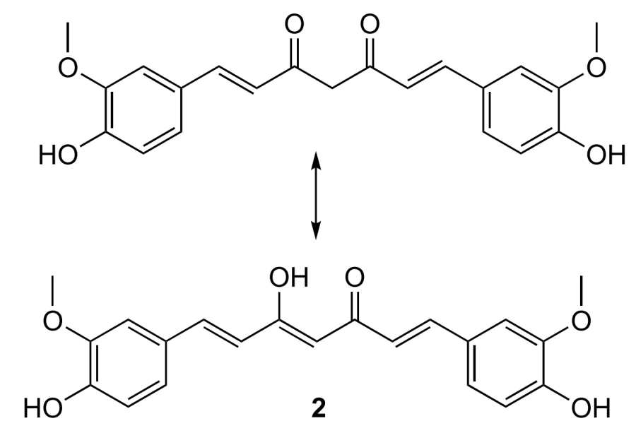
Currently, there are reviews devoted to particular natural additives, for example, extracts of pomegranate Punica granatum L. for the treatment of oral cancer [45], grape seeds for the treatment of oral candidiasis [46], propolis for the treatment of periodontal disease [47], drugs based on curcumin (The antibacterial properties of curcumin are determined by the presence of three active groups in the structure, namely, ketone central groups with keto–enol tautomerism, two double bonds with cis–trans isomerism and phenolic groups (see https://doi.org/10.3390/coatings11050519).) (Structure 2) [48], algae for oral hygiene [49], and extracts of red betel leaves Piper crocatum [50], Azadirachta indica tree [51], Salvadora persica shrub (Salvadoraceae family) [52] and antioxidant secondary metabolites of medicinal plants [53].
It has been described in detail which substances in the extracts have an antimicrobial effect, and probable mechanisms of their interaction with bacterial and fungal cells and their effect on the human body are proposed [50], [51], [52], [53], [54]. These substances include essential oils, flavonoids, alkaloids, terpenes and terpenoids, tannins, polyphenols, phytoncides, carotenoids, etc [18], [50], [55]. However, there are no review works in the literature that evaluate the prospects for the use of natural additives in various fields of dentistry and provide their comparative characteristics, which is important for the use of modified dental materials in clinical practice. This review provides a comparative analysis of such characteristics, recommendations for the use of natural extracts in various fields of dentistry, such as therapeutic dentistry (caries prevention and dental filling materials), endodontics, orthodontics, dental prosthetics and implantology, and also indicates what research is necessary for the implementation of dental materials into clinical practice.
2. Relevance of the research area
According to the Scopus scientometric database, the largest number of publications are devoted to mouthwashes containing natural antimicrobial additives. As can be seen from the histogram (Figure 4.), over the past 10 years, the number of publications devoted to mouthwashes in therapeutic dentistry has amounted to more than 400, with filling materials with antimicrobial action and irrigants for root canal treatment being the second most abundant (more than 100 publications). The smallest number of publications is devoted to toothpastes, dentures and orthodontic appliances and adhesives containing plant extracts (less than 100 publications).
![[{"type":"paragraph","data":{"text":"Number of publications in the field of dental materials over the past 10 years (Scopus database)."}}]](/storage/images/resized/QmPFS6ohOhfmyqkhskPRnqqV1YG5D0Zdy7sXN7qJ_xl.webp)
The greatest publication activity in the field of oral care products was observed in 2019 and 2021. Two waves of
publication activity can be distinguished. The first wave was from 2013 to 2017, and the second one was from 2018 to 2022. In the field of dental filling materials, there was a gradual increase in publication activity, reaching a maximum in 2021 and 2022. The lower interest of researchers in dental filling materials with antimicrobial effects compared to mouthwashes is probably due to the fact that the prevention of dental and periodontal diseases and oral hygiene using mouthwashes is cheaper and easier to tolerate for patients than their treatment.
In turn, the greater interest of researchers in dental filling materials compared to dentures is due to the fact that the use of dental filling materials saves person’s own teeth and does not require the additional use of adhesives and fixation systems for metal or ceramic crowns. In addition, dentures require additional, more thorough care and greater financial costs than filling materials due to the risk of secondary caries.
3. Natural additives for dental filling materials
Glass ionomer cement (GIC) consists of three components: water-soluble polyalkenoic acids, ion-leaching fluoroaluminosilicate glass as a fine powder able to release fluoride ions, and water. GIC is the most common dental filling material because it is biocompatible, quickly cures, has high adhesion to the tooth structure, good colour combination and translucency, and is capable of releasing (Glass ionomer cements are inherently biologically active in part because they release biologically active ions (fluoride, sodium, phosphate and silicate) into the surrounding aqueous media at levels at which they are biologically beneficial.) fluoride, calcium Ca2+, strontium Sr2+, phosphate PO43– and silicate SiO44– ions to the oral cavity, which promotes tooth remineralization [56]. The inclusion of plant extracts into GIC produces dental filling materials that have antibacterial activity against gram-positive sugar-dependent bacteria S. mutans. For being included into GIC, the plant extract is added to distilled water and mixed with GIC component powder. Curing of GIC occurs in two stages. At the first stage, calcium Ca2+, aluminium Al3+ and silicate ions SiO44– are released into an aqueous environment upon contact with the powder. At the second stage, Ca2+ and Al3+ ions cross-link the chains of polyalkenoic acids (for example, polyacrylic acid; copolymer of acrylic, itaconic and maleic acids), while silicate ions form silica gel [57].
Plant extracts can act as both antibacterial additives and photosensitizers, absorbing light at wavelengths between 600 and 800 nm on exposure to a non-thermal light source (laser) and causing the production of ROS, enzymatic changes in DNA and structural modifications in the bacterial cell membrane.
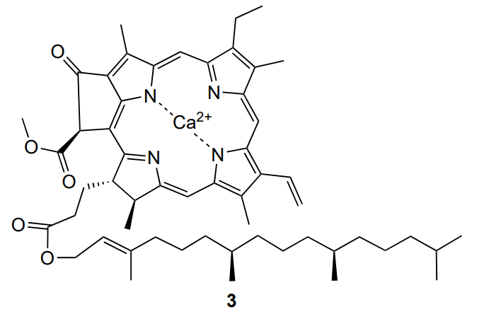
For example, an extract of Dioscorea altissima (Dioscoreaceae family) added to GIC powder contains chlorophyll, which acts as a photosensitizer [58]. As a result, according to the authors, it was possible to improve the antibacterial effect of GIC against S. mutans upon laser irradiation due to the calcium chelation with the porphyrin ring of chlorophyll (3).
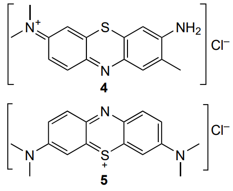
The extract acted as a safe alternative to toxic photosensitizers such as toluidine blue (4) and methylene blue (5).
However, the green colour of the extract has an adverse effect on the aesthetic properties of the dental filling material. For the practical use of this material, additional studies of the chemical composition of the extract and the mechanism of its antimicrobial action are necessary to establish whether it is possible to remove the pigment and whether it affects the antimicrobial activity. Unfortunately, the mechanical and adhesive properties of GIC have not been studied in the cited work.
It was found [59], [60] that ethanol extracts of a mixture of several plants, namely. olive Olea europaea leaves, fig tree Ficus carica, and Salvadora persica leaves and roots, have significant antimicrobial activity against S. mutans (Table 1) and when included in GIC, they do not have a statistically significant effect on the GIC film thickness, compressive strength or shear bond strength, which inhibits the sliding of the filling material along the tooth structure.
![Mean values of inhibition zones (mm) against <i>S. mutans</i> (incubation for 48 hours at a temperature of 37±1 °C)[<a class="anchor" href="#reference-593">60</a>].](/storage/images/resized/JclqhCpUZw1c2fvnoH2dpTpcpvU6Z0wTEzkSRFjj_xl.webp)
According to the data on inhibition zones (see Table 1) measured using a micrometer, the higher the extract content in relation to the water intended for the preparation of GIC, the greater the inhibition zone [60]. This dependence contradicts earlier studies [61], in which the antibacterial effect of extracts did not depend on concentration. Unfortunately, there is no explanation for this fact.
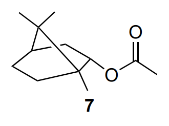
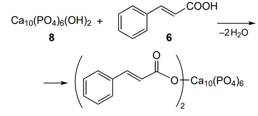
In addition, GIC containing the extract had greater shear bond strength (5.1 MPa) compared to GIC containing chlorhexidine diacetate (1.7 MPa). The authors [59] explain this finding by the presence of cinnamic acid (6) and bornyl acetate (7) in the extract, which improve the release of ions from the GIC surface by lowering the pH and reacting with dentin hydroxyapatite (8) (Scheme 1 shows an example of reaction for cinnamic acid).
The composition of these extracts was determined [60] using gas chromatography–mass spectrometry, which detected the presence of [38] compounds in the extract: monoterpene hydrocarbons and alcohols, sesquiterpenes, coumarins, phenols, flavonoids and saponin. These compounds destroy the bacterial cell membrane (terpenoids) and cause protein denaturation (coumarins and phenols) and leakage of proteins and enzymes of bacterial cells (saponin). Establishing the composition of extracts is a significant advantage of this study. However, the influence of the extract on water absorption, water solubility and flexural strength of GIC, parameters that affect the durability and stability of GIC, remained unexplored. The flexural strength is a more important parameter than the compressive strength, since it is sensitive to small changes in the structure of the material and may simulate a clinical loading situation.
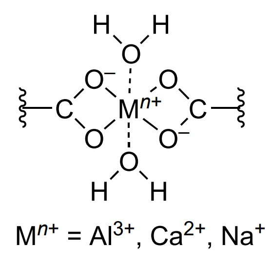
The same research group investigated the effect of a mixed ethanol extract of Olea europaea, Ficus carica and Salvadora persica plants on water absorption, water solubility and flexural strength of GIC [62]. The authors found that the extracts did not have a statistically significant effect on the water absorption and water solubility of GIC, but increased the flexural strength from 11.8 to 26.1 MPa at a mass ratio of extract to water of 2 : 1. The flexural strength was higher than that of chlorhexidine-modified GIC (15.3 MPa). The authors attributed the increase in the flexural strength to the presence of unreacted powder particles in the matrix, (The extract can influence the amount of unreacted powder particles in the matrix, which act as reinforcing fillers, preventing the propagation of cracks within the cement [62].) which prevented the propagation of cracks, and to an increase in the degree of cross-linking due to the formation of polysalt bridges (Scheme 2) with participation of cinnamic acid and bornyl acetate. However, the degree of cross-linking was not determined in the study, and from the chemical point of view this assumption is difficult to substantiate, since cinnamic acid is monocarboxylic and, on the contrary, it is expected to reduce the degree of cross-linking, while boronyl acetate (7) does not show acidic properties at all. It should also be noted that dynamic forces were not taken into account when measuring the strength of the modified GIC, nor were factors such as clinical conditions, component mixing technique, GIC manufacturer, ratio of glass powder to the cement preparation liquid, and water content.
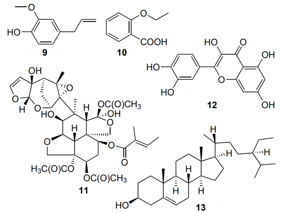
Since the disadvantages of GIC as a filling material are its high cost and sensitivity to restoration techniques, new filling materials have been developed based on hydrogenated rosin, eugenol (9) and 2-ethoxybenzoic acid (10) as binding agents, and zinc oxide, aluminium oxide and sodium fluoride as a cement base. To endow the material with the necessary antibacterial activity against S. mutans, an ethanol extract of the Azadirachta indica neem tree was proposed [63]. The authors explained the choice of ethanol rather than water for extraction by additional antimicrobial activity of ethanol and better solubility of extracted phenols in ethanol. A study by diffusion method in agar showed the presence of inhibition zone of 11.8 mm with the addition of 6 wt.% extract. This value is comparable with that attained by the addition of 5 wt.% chlorhexidine diacetate/cetrimide mixtures (12 mm) [64]. Interestingly, extracts of Azadirachta indica twigs without bark showed lower MIC values (3.125%) compared to leaves (6.25%) or twigs with bark (50%). However, the authors [63] gave no explanation to this fact. In our opinion, the leaf extract contains larger amounts of azadirachtin (11), quercetin (12) and β-sitosterol (13) than the bark, which determines better antimicrobial effect of the leaves compared to the bark [65]. In addition, to explain the better effect of the extract of branches compared to leaves, it is necessary to compare the contents of components in them depending on the place and conditions of plant growth, the time of the year when the leaves and branches were collected, ambient temperature, the presence of moisture, etc.
Mathew and Sghaireen[66] compared the antibacterial effect of cements containing extracts of leaves, stems and fruits of the Christ’s thorn jujube (Ziziphus spina-christi) against S. mutans bacterium. As a result, the greatest antibacterial effect was observed for leaves (inhibition zone diameter (IZD) of 75±5 mm) compared to stems (IZD 66±5 mm) and fruits (IZD 58±7 mm), which was attributed to high content of polyphenols and saponin in the leaves.
A disadvantage of cements containing Ziziphus spina-christi extract compared to cements containing the antibiotic gentamicin is their higher MIC because unlike gentamicin as a single substance, the extract is a mixture of bioactive substances.
In the cited studies[63], [66], there is no comparison of the inhibition zones with the control group (unmodified composite). On the contrary, Paulraj and Nagar[67] compared the antibacterial effects against S. mutans for cement powders containing an aqueous extract of Terminalia trees (Terminalia chebula, Terminalia belerica) and Indian gooseberry Phyllanthus embelica (Triphala Ayurvedic drug) (Triphala consists of three plants: Terminalia chebula, Terminalia belerica, Phyllanthus embelica.) with that of unmodified cement. The average inhibition zone for the modified cement was 11.5 mm, while the unmodified cement also had little antibacterial activity (5.5 mm), which is likely due to the release of fluoride from the cement. However, only 10 ppm of fluoride is released, which cannot produce the required antibacterial effect. The authors [67] attributed the antibacterial effect of the aqueous extract of Triphala to tannic acid (14), which is adsorbed on the surface of S. mutans cells, inducing protein denaturation and cell death.
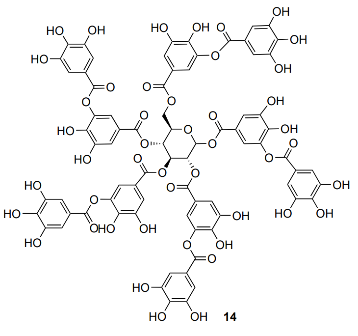
A cement containing an ethanol extract of propolis also showed a similar antibacterial effect (inhibition zone diameter of 12 mm), due to galangin (15), caffeic acid (16) and flavonoids, which change the permeability of the bacterial cell wall and inhibit bacterial enzymes [67].
However, additional research is needed to elucidate the effect of Ziziphus spina-christi, Triphala and propolis extracts on the physicochemical and mechanical properties of dental filling materials for their implementation in clinical practice.
Dental filling materials must be resistant to acid erosion, show chemical adhesion to tooth enamel (about 6–7 MPa) and dentin (about 4–5 MPa), hardness, radiopacity, wear resistance, and good appearance. To use GIC modified with plant extracts, dentists need to further investigate these parameters, conduct sanitary and chemical studies by determining the content of fluoride ions in artificial saliva and plasma, and also study how the biologically active substances of the extracts would interactwith GIC components, which is of great importance for analysis of curing. IIf GIC is cured not only chemically, but under the action of both light and chemicals, then the cement contains oligomers and light polymerization initiators and additional research is necessary to elucidate the effect of the modifying additive on the radical polymerization, depending on the degree of cure.
4. Natural additives in endodontics
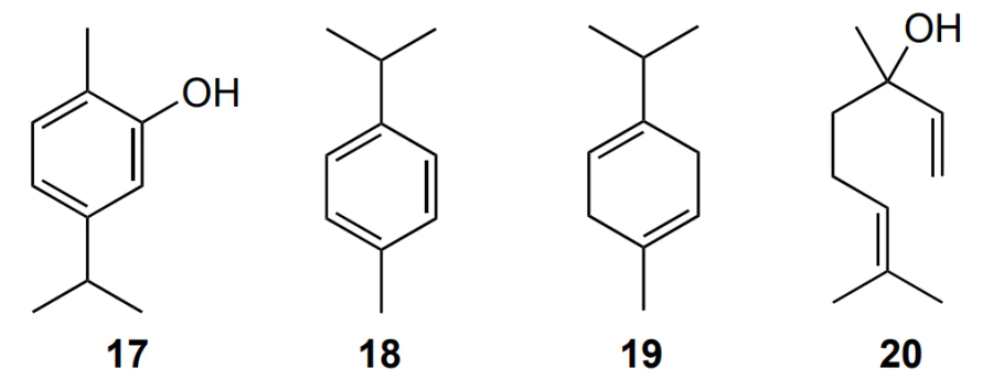
Non-synthetic root canal irrigants, in particular plant essential oils of thyme Thymus lamiaceae and oregano Origanum vulgare (Lamiaceae family) [68], extracts of suran Amorphophallus paeoniifolius and aravi Colocasia esculenta [69] and extract of Moringa oleifera [70] showed activity against root canal microflora, represented mainly by gram-positive bacteria E. faecalis, causing apical periodontitis and secondary root infections. The antibacterial effect of thyme and oregano extracts is due to the biologically active molecules they contain, including thymol (1), carvacrol (17), p-cymene (18), γ-terpinene (19) and linalool (20).
Unlike lemongrass and melaleuca essential oils, these extracts stand out regarding their MIC values against E. faecalis. [68], [69], Table 2 shows the MIC values for the ndicated extracts.
Unfortunately, there is no explanation for the higher MIC value of oregano extract compared to thyme in these studies. In our opinion, this is due to the higher content of thymol 1 in the thyme essential oil extract (approximately 55%) than in oregano extract (approximately 15%) [71].
An aqueous ethanol extract of leaves of the guava tree Psidium guajava L. (Myrtaceae family) showed a lower MIC value against the bacteria E. faecalis than sodium hypochlorite or oregano and thyme extracts [72]. This value was lower than that of the commercial irrigant, sodium hypochlorite, which makes the extract promising as an irrigant. Abreu Baldoni et al [72]. attributed the antibacterial effect of the extract to the action of secondary metabolites identified during phytochemical analysis, such as phenolic compounds, tannins, terpenoids, flavonoids and glycosides.
However, the studies cited above do not compare the efficacy of plant extracts with that of the commercial irrigant chlorhexidine, long-acting calcium hydroxide-based agents or with the antimicrobial action of the diode laser, which can provide access to poorly accessible areas of the root canals. (The root canal is treated with a diode laser (0.5–7 W, 940 nm), which delivers energy through thin flexible fibres to the root canal.)
The extracts of suran Amorphophallus paeoniifolius and aravi Colocasia esculenta are better studied in this regard. Their antibacterial effect did not differ from that of calcium hydroxide and was due to the saponins they contained, which reduce the permeability of the bacterial cell and inhibit its growth [69]. However, the number of colony forming units (CFU) was higher when plant extracts were used than in the case of chlorhexidine gel or diode laser irradiation. The survival rate of E. faecalis bacteria varied in the following order: 2% chlorhexidine gel < diode laser irradiation < suran extract < aravi extract < calcium hydroxide (
![Bacterial survival rate upon intracanal sampling 7 days after of root canal treatment[<a class="anchor" href="#reference-602">69</a>].](/storage/images/resized/Z3M4dsbjDTruaoaSyUpsKk6I7mlNS2WOvAewmsZJ_xl.webp)
The authors gave no explanation to the higher antibacterial activity of chlorhexidine compared to plant extracts. This is probably due to the ability of chlorhexidine to penetrate into the bacterial cell with subsequent leakage of the cell contents — adenosine triphosphate and nucleic acids. In addition, chlorhexidine binds to hydroxyapatite and soft tissue, thus reducing the adhesion of microorganisms. The saponins in the plant extracts of suran and aravi, despite the fact that they destroy the cell wall, cannot bind to hydroxyapatite, which makes them less effective.
An advantage of natural plant extracts over chemical irrigants (sodium hypochlorite) include the lack of toxicity since they do not release chlorine and dissolve only necrotic pulp residues rather than vital pulp, do not irritate periapical tissues and do not reducte dentin flexural strength; they do not require heating to 60 °C to enhance the solubility (Given the toxic potential of sodium hypochlorite, it would be inappropriate to use a solution at a high concentration. Increasing the temperature of sodium hypochlorite at low concentrations has been suggested as an attempt to resolve this limitation, as heating enhances the dissolution and antibacterial properties of the irrigant.) and to improve the antibacterial effect [73], [74], [75]. Unlike synthetic irrigants [76], natural irrigants do not irritate the skin and do not have a cytotoxic effect on human osteoblasts. However, the antibacterial effect of decoction of Moringa oleifera leaves is less pronounced than that of sodium hypochlorite, as evidenced by smaller inhibition zones against E. faecalis and S. mutans (Figure 5). [70]
![[{"id":"tKngAteUvb","type":"paragraph","data":{"text":"Inhibition zones of the <i>E. faecalis</i> (blue columns) and <i>S. mutans</i> (red columns) bacteria. The samples were prepared using extracts of appropriate weights of dried <i>M. oleifera</i> leaves (2.5 and 5 wt.%) and, separately, a 5% aqueous solution of sodium hypochlorite. The Figure was created by the authors using published data[[ type=\"anchor\" referenceId=\"603\" ]]."}}]](/storage/images/resized/XkFxydUSQBJKZcu9NgzuyhzMswhzQs9E9S6P8SoQ_xl.webp)
The advantage of Moringa oleifera over sodium hypochlorite is its higher efficiency in removing the smear layer on dentin. According to Natsir et al. [70], this is due to the presence of flavonoids in the decoction. Flavonoids decompose hydroxyapatite, which is a component of the smear layer, and release calcium ions Ca2+ and hydrogen phosphate anions HPO42–, which promote remineralization.
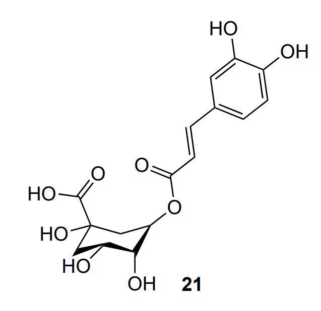
A drawback of the decoction of Moringa oleifera leaves is lower diameter of the inhibition zone compared to that for the extract of the herbaceous plant Epilobium parviflorum Schreb (Onagraceae family). For an aqueous ethanol extract of Epilobium parviflorum Schreb, Șachir et al [77]. found an inhibition zone of 18–20 mm against S. mutans and 11.5–12.5 mm against E. faecalis. In our opinion, this factor is due to the high content of flavonoids in the Epilobium parviflorum Schreb extract compared to the Moringa oleifera extract and the presence of polyphenols in large quantities in the extract (10.45 equiv. Of chlorogenic acid (21) per gram of vegetable powder polyphenols).
An interesting fact established by Șachir et al. [77] is that aqueous extracts of Epilobium parviflorum Schreb did not have an antibacterial effect against S. mutans, E. faecalis, S. mitis, white Staphylococcus, or S. sanguis, and it is necessary to use aqueous ethanol extracts. The authors explained this by better ethanol solubility of the polyphenols contained in the extract. To improve the solubility of polyphenols and flavonoids, it was proposed to carry out extraction at elevated temperatures (40 °C).
Unlike the aqueous extract of Epilobium parviflorum Schreb, the aqueous extract of Lawsonia inermis (Lythraceae family) showed an antibacterial effect against E. faecalis with an inhibition zone of 11 mm [78]. The antibacterial effect of the extract is due to secondary metabolites and their active components, such as lawsone (2-hydroxy-1,4-naphthoquinone) (22), mannitol (23), gallic acid (24) and tannic acid (14).
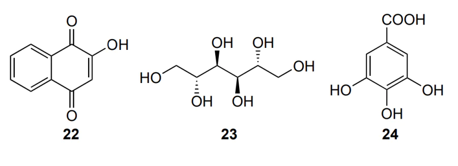
The mechanism of the antibacterial action of lawsone (13) is probably related to increased formation of ROS and damage to protein and DNA macromolecules and bacterial cell lipids (Figure 6) [79]. However, the inhibition zone of the extract was 3 mm smaller than that of the commercial irrigant chlorhexidine digluconate. The reason for this fact was not explained by the authors.
![[{"id":"mVhthvSN7x","type":"paragraph","data":{"text":"Mechanism of the antibacterial action of lawsone (using the example of <i>E. faecalis</i>)."}}]](/storage/images/resized/d6l4LwbCD57xDNq88uBxKLjT9jCMWaxSZNlcbFiE_xl.webp)
In addition to E. faecalis bacteria, C. albicans fingi are also found in infected root canals and account for 3–18% of the total amount of pathogens [72]. The number o studies devoted to the effect of extracts on C. albicans fungi is much smaller compared to the number of papers considering E. faecalis bacteria, being less than 20 publications over the past 10 years. Some of the extracts, for example, aqueous and ethanolic extract of Euclea divinorum Hiern (Ebenaceae family) [80] and ethanolic extract of Curcuma Longa [81] inhibited the growth of E. faecalis, but showed low activity against C. albicans fungi. It should be noted that, like in the study by Elizabeth et al. [78], ethanolic extracts were more effective than aqueous ones. Maina and Dimba [80] attributed this fact to the activity of the polyphenol oxidase enzyme in the aqueous extract, which destroys biologically active polyphenols and reduces the antibacterial activity.
Basheer and Sharma [82] measured the inhibition zones for both E. faecalis bacteria and C. albicans fungi under the action of an ethanolic extract of two plants: black thymine Nigella sativa (Ranunculaceae family) and carnation Eugenia caryophyllus (Myrtaceae family). It was found that the plant extract had better antimicrobial activity compared to 2.5 wt.% sodium hypochlorite and 2 wt.% chlorhexidine solution, as evidenced by the large diameters of the inhibition zones (Figure 7). In addition, a synergistic effect of the resulting extract comparable with the action of separate extracts was observed. The viability of L929 fibroblast cells was more than 99%, indicating the biocompatibility of the extract.
![[{"id":"uOPzBjuYIC","type":"paragraph","data":{"text":"Inhibition zones of <i>E. faecalis</i> and <i>C. albicans</i> bacteria. The Figure was created by the authors using published data[[ type=\"anchor\" referenceId=\"615\" ]]."}}]](/storage/images/resized/GrSX6oG5UnzfHjdUCmeCCioR143QOA70wUdjOCfi_xl.webp)
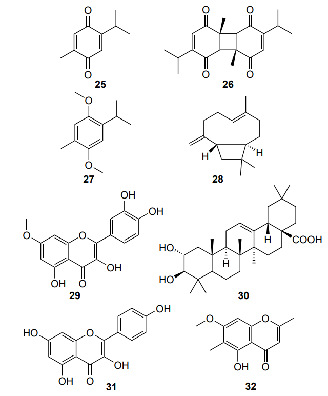
The authors attributed the good antimicrobial effect to the presence of numerous pharmacologically active substances in the extract, such asthymoquinone (25), dithymoquinone (26), thymol (1), thymohydroquinone (27), eugenol (9), β-caryophyllene (28), rhamnetin (29), maslinic acid (30), kaempferol (31) and eugenitin (32). These substances are capable of separating cell membrane lipids, thus inducing cell lysis.
Unlike the authors cited above, which focused on antibacterial effects against single microorganisms, Yuanita et al. [83] examined the effect of cocoa pod husk extract on the E. faecalis biofilm. The results showed that the addition of 6.25 vol.% extract reduced the E. faecalis biofilm thickness; this was the threshold value: a lower content of the extract did not have a statistically significant effect on the biofilm thickness (Table 4). The authors explained the decrease in the biofilm thickness by the action of alkaloids, flavonoids, tannin and saponin contained in the extract.
In addition to E. faecalis bacteria, the root canal biofilm also contains other microorganisms such as C. albicans fungi, S. gordonii and S. mutans bacteria, etc. Wanicharat et al. [84] studied the effect of the extract of the fruit Boehmeria macrophylla (Urticaceae family) kernels on the growth of a multispecies biofilm consisting of E. faecalis, C. albicans and S. gordonii. The authors compared the effect of the extract with those of calcium hydroxide and 2 wt.% chlorhexidine gel. The biofilm treated with 50 mg mL–1 (see j) plant extract had a higher proportion of dead cells than calcium hydroxide- and chlorhexidine-treated biofilms.
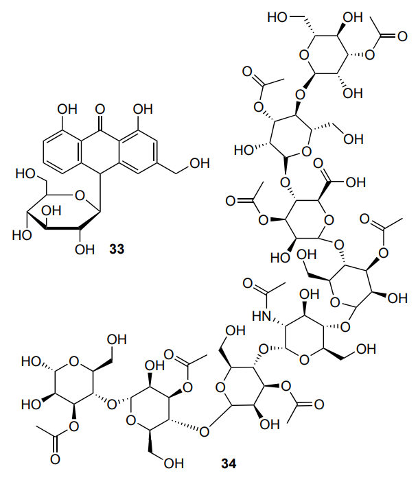
An aqueous extract of Aloe vera (Asphodelaceae family) was able to inhibit the growth of C. albicans fungal biofilm within 24 hours of incubation [85], with the antifungal effect being higher than that of a commercial irrigant, a 17 wt.% EDTA solution. However, a higher Aloe vera concentration (75 wt.%) was required for the inhibition. The authors attributed the antifungal effect of the extract to the action of compounds such as aloin (33) and acemannan (34), which interact with the fungal cell walls.
However, irrigants cannot eliminate microorganisms from root canals with 100% efficiency. A promising replacement for irrigants is photodynamic therapy using non-toxic curcumin 2 and curcuminoids as photosensitizing dyes or a combination of the antimicrobial photodynamic therapy with shock wave enhance emission photoacoustic streaming (SWEEPS). The SWEEPS method uses an erbium-doped yttrium aluminum garnet (Y3Al5O12) laser to generate a flow of bubbles enhanced by shock waves. Ensafi et al. [86] conducted a study on E. faecalis biofilm in the root canals of extracted human teeth and found that a combination of photodynamic therapy using solutions of curcumin 2 and nanocurcumin in dimethyl sulfoxide, irradiation with a light-emitting diode at 390–480 nm and SWEEPSk is more effective than the photodynamic therapy alone. This is due to the removal of microorganisms from hard-to-reach areas of the root canals. Nanocurcumin under the brand name Curcuden was used to improve the bioavailability of curcumin 2.
In the field of endodontics, mouthwashes composed of oil-inwater microemulsions of curcumin 2 can also be used in combination with photodynamic therapy, since curcumin 2 is a hydrophobic compound.
However, despite its low toxicity to human skin fibroblasts [87], curcumin 2 is hydrolytically unstable and stains the tooth enamel [88], and its antibacterial activity against E. faecalis is worse than that of 5 wt.% sodium hypochlorite [89].
Despite the well-studied antimicrobial effect of plant extracts against root canal microorganisms, their use in endodontics requires additional research on the wetting ability of irrigants, foam formation, dissolution of the organic matrix of root canal dentin, removal of dentinal sawdust and organic residues from the root canal system after instrumental treatment in the course of therapy and the effect of irrigants on periapical tissues and determination of the optimal time for the endodontic irrigant to remain in the root canal. Furthermore, the combined treatment of the smear layer on the dentin surface with solutions of commercial irrigants could ensure its complete removal.
5. Natural additives in oral care products (therapeutic dentistry)
Mouthwashes containing plant extracts can be used not only in endodontics, but also in therapeutic dentistry for the prevention of carious lesions, periodontitis, and gingivitis.
For example, a mouthwash based on water-soluble fractions of a combined extract of the oak Quercus infectoria (Fagaceae family) and the perennial herb Scrophularia striata (Norichinaceae family) has antimicrobial activity against cariogenic microorganisms S. mutans, S. sobrinus and C. albicans [90]. The liquid was prepared by dissolving the fractions in a mixture of propylene glycol, ethanol and water (10:10:80 v/v/v), with propylene glycol acting as a preservative and providing a sweet taste to the liquid. To make the liquid pH neutral and eliminate the adverse effects on tooth enamel of the acidic medium caused by acids present in the extracts (The extract contains tannic acid pH = 3.5), sodium bicarbonate was added. The S. mutans bacteria were most sensitive to the action of the extract, and the C. albicans fungi were least sensitive. According to the authors, this was due to the fact that the active components of the extract (mainly tannic acid 14) require more time to penetrate the cell wall of the fungus compared to the bacterial cell. The galloyl group of tannic acid 14 binds more easily to the peptidoglycan layer of the bacterial cell wall and destroys the cell wall than to the chitin of the fungal cell wall.
A study of the stability of the resulting mouthwash at 25 °C for 60 days showed the absence of statistically significant changes in the colour, odour, taste, and transparency. However, the antimicrobial properties of the liquid deteriorated, which, according to the authors, was due to the oxidation of decomposition products of tannic acid 14 (Tannic acid is a hydrolysable tannin that is degraded to more hydrophilic compounds such as glucose, gallic acid, pyrogallol and
oxalic acid by successive hydrolysis, decarboxylation and oxidation. Some of the decomposition products of tannic acid are biologically active. Studies have shown that heat treatment partially hydrolyses tannic acid to more potent antibacterial and antioxidant compounds. We hypothesize that the reduced antimicrobial activity of stored mouthwash samples may be due to further oxidation of the active compounds to quinine by a reaction that is accelerated in neutral and alkaline solutions.) to give substances devoid of antimicrobial activity. Unfortunately, no data of toxicological studies are given in the paper.
Zulkamal et al.[91] studied an aqueous extract of the butterfly pea Clitoria ternatea (Fabaceae family) not only for antibacterial effect, but also for the toxicity to shrimp. The extract was non-toxic, as the lethal concentration LC50 was higher than 1000 µg mL–1.
The aqueous extract destroyed the S. mutans bacterial cells within 36 hours of treatment due to the presence of alkaloids, tannins, glycosides, anthocyanins and flavonoids, which was confirmed by SEM micrographs (Figure 8) [91]. The micrograph shows cellular debris and irregular shape of bacterial cells due to lysis of the contents (Figure 8b).
![[{"id":"hDNlIMSLhU","type":"paragraph","data":{"text":"SEM images of <i>S. mutans</i> treated with <i>Clitoria ternatea</i> aqueous extract at a concentration of 400 μg mL<sup>–1</sup>. (a) 0 hours (control) and (b) 48 hours[[ type=\"anchor\" referenceId=\"623\" ]]."}}]](/storage/images/resized/BuBbEoEakd0VBm6X1xLJeTxI9cjGBw14aXdqXn79_xl.webp)
The antibacterial effect of extracts was compared to that of a commercial product, a 0.2 wt.% solution of chlorhexidine digluconate [92], [93]. Nasution and Syahputra [92] reported that a mouthwash based on Zanthoxylum achantopodium, glycerol, sorbitol (to give a sweet taste), sodium carboxymethylcellulose (to ensure the required viscosity) and water showed a more pronounced antibacterial effect against S. sanguinis bacteria, which are involved in the formation of dental plaque and development of periodontitis, than 0.2 wt.% chlorhexidine gluconate. The average numbers of bacteria (CFU/mL) for the developed liquid containing 8 wt.% of the extract and chlorhexidine were 204.33±108.45 and 460.67±137.59, respectively. The authors explained this fact by the action of secondary metabolites, alkaloids, flavonoids, glycosides, saponins, tannins and terpenoids, present in the extract. Alkaloids inhibit nucleic acids and pump out proteins from the bacterial cell; flavonoids form complex compounds with extracellular proteins; saponins increase the permeability of the cell membrane and inactivate enzymes; tannins damage the genetic material of the bacterial cell and inhibit enzymes; and terpenoids form bonds with porins, thereby inhibiting the transport of nutrients into the cell. According to the authors, the developed mouthwash, unlike chlorhexidine, would not change the colour of teeth or taste sensations and would not cause drying and burning in the mouth and desquamative skin lesions.
Conversely, Naghsh et al. [93] found 0.2 wt.% chlorhexidine digluconate to be more effective against the S. sanguinis bacteria than a Matrika chamomile extract or rinses based on Aloe vera (Aloaceae family) containing anthraquinones, phenolic compounds, saponin, tannin and organic acids as the active components. Unlike mouthwashes with herbal ingredients, chlorhexidine reduced the number of bacterial colonies to zero.
In addition, the inhibition zones were small (less than 5 mm), which does not allow the use of extracts as mouthwashes in therapeutic dentistry for the prevention of caries and periodontal diseases. In addition to the prevention of caries and periodontal diseases, mouthwashes based on plant extracts can be used to heal wounds and oral ulcers. For example, Monica et al. [94] found that an aqueous extract of a mixture of Cinnamomum verum (Lauraceae family), Coleus aromaticus/amboinicus (Lamiaceae family) and Mentha piperita (Lamiaceae family) has anti-inflammatory and antimicrobial effects against pathogenic wound microbes P. gingivalis and S. aureus. The authors attributed the anti-inflammatory effect of the herbal composition to the inhibition of inducible nitric oxide synthesis and cycloxygenase-2 expression in the central nervous system under the action of 2-hydroxycinnamaldehyde present in cinnamon. However, the antibacterial effect of the extract was worse than that of the antibiotic ampicillin, as evidenced by the smaller diameter of the inhibition zone (15–18 mm for the extract and 20–21 mm for ampicillin). The reasons for this are yet to be explained.
Apart from homogenous solutions, nanoemulsions are also used as mouthwashes. Compared to solutions, nanoemulsions have optical transparency and stability. In particular, Nehavarshini et al. [95] prepared an oil-in-water nanoemulsion based on hot gingelly oil, hot neem oil, polysorbate 20 as a nonionic surfactant, and ultrapure water. The nanoemulsion was mixed with a plant extract based on Acacia arabica trees (Fabaceae family), black myrobalan Terminalia chebula (Combretaceae family), Terminalia bellerica (Combretaceae family) and Emblica officinalis (Philantaceae family) to form a nanoemulsion mouthwash. The antibacterial properties of the nanoemulsion with a plant extract were studied against S. mutans, lactic acid bacteria L. casei and A. viscosus, which cause periodontal diseases and bad breath. The nanoemulsion liquid inhibited the adhesion of S. mutans bacteria by 73%, L. casei bacteria by 63%, and A. viscosus bacteria by 61%, which was higher than these values for chlorhexidine. In addition, the nanoemulsion liquid not only inhibited the adhesion of microorganisms, but also inhibited the growth of biofilm, prevented caries in artificial mouth assay (Artificial mouth assay is a system of multi-station, continuous-flow chambers for culturing microflora that mimic the biological and physiological activity seen in the oral cavity.) and promoted tooth remineralization. The authors attributed the antibacterial and antibiofilm effect of the nanoemulsion liquid to the destruction of the lipid membrane of the bacterial cell.
The publications discussed above [94], [95] describe in vitro studies. Kiani et al. [96] reported the use of a mouthwash containing a propolis extract by patients with gingivitis aged 19 to 55 years with at least 20 teeth. The authors found a decrease in the plaque index and papillary bleeding index compared to the control group after two weeks and after a month of observation, with no statistically significant change in the tooth colour being observed. There were also no possible side effects observed, such as reddening of the gums and mucositis (inflammation of the tissues of the oral cavity). Thus, propolis extract has a wound healing and therapeutic effect in patients with gingivitis. However, the study included only 32 participants (16 persons in both the test and placebo groups). In the future, the authors plan to conduct a study with a larger sample size, which will allow the use of a propolis-based mouthwash for the treatment of gingivitis.
Kakoei et al. [97] conducted a clinical study including 40 patients. The authors studied the effectiveness of mouthwash based on 2% aqueous extract of henna Lawsonia inermis (Lythraceae family), 2% ethanol, 10% glycerol and 0.1% methylparaben solution and a commercial mouthwash based on 0.2% chlorhexidine solution as adjunctive therapy for oral lichen planus. As a result, the authors found that the henna extract liquid and chlorhexidine were equally effective in reducing pain intensity as assessed on a visual analog scale, after two weeks of the study. The wound-healing effect of henna extract, according to the authors, is due to the presence of coumarins, flavonoids, tannins, naphthoquinones, terpenes, steroids and alkaloids. However, a longer study in a larger number of patients is required to evaluate the therapeutic effects of the henna extract liquid and study the side effects, for example, in people with deficiency of the enzyme glucose-6-phosphate dehydrogenase.
Unlike the above-cited authors [96], [97] Kim and Nam [98] used a randomized placebo-controlled clinical trial of 64 patients who were assigned to a saline solution and Sambucus williamsii var. coreana. In addition, the authors [98] studied the antimicrobial effect of a mouthwash. As a result, the extract was found to reduce clinical indicators associated with periodontal disease, namely, the O’Leary index, plaque index and gingival index after 5 days of observation, while the saline solution did not show a statistically significant difference. A study of the antimicrobial effect of the extract showed a decrease in the number of gram-positive P. micra and gram-negative T. denticola, P. intermedia and E. corrodens, P. gingivalis bacteria, associated with periodontal disease in the subgingival dental plaque of the mandible, after 5 days of observation (Figure 9).
![[{"id":"a6BYE1pbcK","type":"paragraph","data":{"text":"A randomized, placebo-controlled clinical trial evaluating the use of a mouthwash containing the <i>Sambucus williamsii</i> <i>var. coreana</i> extract for prevention of gingivitits (blue column is the baseline, red column is after 5 days). The Figure was created by the authors using published data[[ type=\"anchor\" referenceId=\"631\" ]]."}}]](/storage/images/resized/1fk8kHQ61kRRiti9er5bY8OB3r7VNQKlvaOoIOZM_xl.webp)
The authors note that the extract was more effective for rinsing the mandible than the maxilla because the mandible was in a better contact with the rinse. According to the authors, for maximum effect, it is necessary to treat the entire mouth cavity when rinsing, rather than simply hold the liquid in the mouth. These results indicate good prospects for using Sambucus williamsii var. coreana extract for the prevention of gingivitis.
Kim and Nam [99] found that a mouthwash based on an ethanol extract of Sambucus williamsii var. coreana (concentration 10 mg mL–1) reduces the number of bacteria S. mutans and S. sobrinus in the subgingival dental plaque of the maxilla and mandible after 5 days of observation.
The same research group conducted a clinical study of a mouthwash containing an extract of Lespedeza cuneata (Fabaceae family) and evaluated its antibacterial effect against caries-causing bacteria S. mutans and S. sobrinus [100]. The authors showed a low risk of dental caries based on the Cariview caries activity test and decreasing amount of S. mutans and S. sobrinus bacteria in the subgingival microbiome of the maxilla and mandible after 5 days of follow-up. The total number of subjects was 82 patients, which was greater than in the above studies [98], [99]. The resulting liquid, according to the authors, can be used in therapeutic dentistry to prevent caries and its progression.
Despite the well-studied mechanisms of action and available clinical studies of mouthwashes in the form of solutions, nanoemulsions based on extracts of natural origin need to be studied for their haemostatic and anti-inflammatory effects, their effect on the functions of connective tissue cells, endothelial cells, macrophages and leukocytes, adverse negative reactions for their use in complex therapy of periodontal diseases. In addition, clinical use requires longer utilization of rinses, for at least two weeks.
6. Natural additives in orthodontics
After fixation of orthodontic appliances, a pathological reaction in the surrounding periodontal tissues to the accumulation of oral microorganisms or dental plaque may occur. To prevent inflammation, antimicrobial additives of natural origin are introduced into orthodontic adhesives and orthodontic lightcuring acrylic resins, in particular extracts of cinnamon Cinnamomum verum (Lauraceae family) [101] and extracts of marine green algae Ulva lactuca. [102] Moreover, the physicomechanical properties of the light-curing acrylic orthodontic adhesive Transbond XT, such as shear bond strength and adhesive remnant index, which evaluates adhesion between the orthodontic device and the enamel, were not deteriorated upon the addition of extracts and remained at the level required
for clinical use [101].
The antibacterial effect of cinnamon extracts on grampositive S. mutans bacteria is due to the presence of active components such as cinnamaldehyde (35) and cinnamic acid (6). They reduce the expression of the gtfB, gtfC, gtfD, and gbpB genes involved in the intracellular synthesis of polysaccharides, and the ldh and relA genes encoding the synthesis of lactic acid and guanosine tetra (penta)-phosphate synthetase/hydrolase. In addition, cinnamaldehyde (35) and cinnamic acid (6) prevent tooth demineralization by inhibiting acid-producing enzymes [103]. However, despite being cheap and easy to obtain, plant extracts of cinnamon in orthodontic adhesives showed smaller inhibition zones against S. mutans bacteria compared to the use of titanium dioxide nanoparticles at the same content in the adhesive, equal to 1 wt.% [101].
Extracts of marine green algae Ulva lactuca with concentrations of 0.5%, 1%, 2.5%, 5% and 10% in acrylic lightcuring orthodontic resin system Ortho-Jet for the manufacture of orthodontic appliances reduce the number of S. mutans bacterial colonies by approximately 41%, 61%, 78%, 85% and 95%, respectively, due to the presence of bioactive chlorophyll compounds (type a and b), carotenoids and phycobiliproteins. However, when their concentration in the resin is more than 1 wt.%, the flexural strength of the final products decreases.[102]
An alternative to antibacterial orthodontic adhesives are toothpastes and mouthwashes with natural additives intended for oral care in patients with orthodontic appliances. Such additives include extracts of miswak leaves [104] and turmeric leaves [105], Azadirachta indica tree extract [106] and essential oil of the tea tree Melaleuca alternifolia (Myrtaceae family) [107].
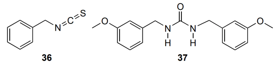
The antibacterial effect of miswak extracts is due to the released sulfur-containing compounds, for example, benzyl isothiocyanate (36), and salvadourea (1,3-bis[(3-methoxyphenyl)methyl]urea) (37), β-sitosterol (13), and flavonoids. However, only methanol extracts of miswak leaves with a MIC of 10 mg mL–1 inhibited the growth of S. mutans bacterial biofilms, while aqueous extracts showed no antibacterial effect[104]. Meanwhile, methanol is poisonous, and using methanol to prepare an extract can cause side effects associated with irritation of soft tissues. Some studies have reported a possible link between methanol and oral cancer [108].
In contrast to miswak leaf extracts, aqueous extracts of the Japanese apricot Fructus mume have an antibacterial effect against S. mutans bacterial biofilms that colonize orthodontic appliances and reduce the viability of bacterial cells by approximately 80% [109]. Unlike turmeric extract [105], which reduces S. mutans in orthodontic patients, and the commercial antiseptic chlorhexidine digluconate, Fructus mume extract does not change the colour of teeth or tongue and taste. The disadvantage of Fructus mume extract include its high MIC (1 g mL–1) compared to chlorhexidine digluconate (250 mg mL–1) and the acidic medium generated by the organic acids present in the extract, which can cause demineralization of enamel with prolonged exposure.
Excellent alternatives to Fructus mume are mouthwashes containing Azadirachta indica neem extract [106] and dental gels containing Melaleuca alternifolia essential oils. Rinse products reduce the mean plaque index and the number of S. mutans microorganisms in areas adjacent to braces, while not causing enamel demineralization [106], and the gels are similar in taste and smell to the commercial Colgate Total gel [107].
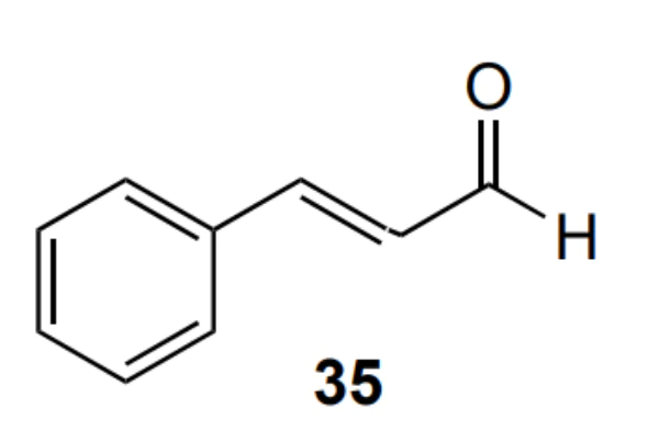
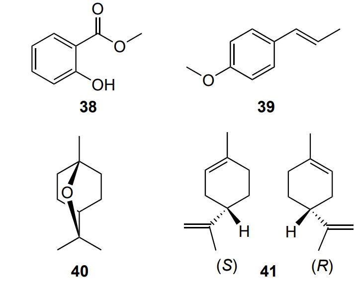
In addition to mouthwashes and gels, there are reported materials for the manufacture of orthodontic appliances (aligners) based on cellulose with biologically active molecules isolated from essential oils adsorbed on their surface and orthodontic nickel titanium arches treated with biologically active molecules [110]. Cinnamaldehyde (35), methyl salicylate (38), trans-anethole (39), eucalyptol (40), limonene (41) and eugenol (9) were used as biologically active molecules,111 because they have low toxicity to human gingival fibroblasts and do not cause contact dermatitis. These compounds inhibited the growth of biofilms of S. mutans and S. mitis bacteria on the surface of the aligners by increasing the hydrophobicity of the bacterial cell surface and reducing their ability to aggregate. In addition, the additives did not deteriorate the mechanical properties (flexural strength) and stability of cellulose-based aligners, which makes their use in orthodontics promising [111].
Eugenol (9) obtained by hydrodistillation of S. aromaticum and diluted with dimethyl sulfoxide showed good antibacterial activity against S. mutans bacteria on the surface of orthodontic nickel–titanium wires, as evidenced by an inhibition zone of more than 20 mm [110]. However, unlike cinnamaldehyde (35), eugenol (9) causes allergic contact dermatitis, which limits its use in orthodontics [112].
In addition to biologically active substances, the plant mixture Padma hepaten based on chebulic myrobalan, amla fruits, and belliric myrobalan is used in from Tibetan medicine. The mixture inhibits the growth of S. mutans bacterial biofilm on the surface of orthodontic appliances made of polyvinyl chloride. This mixture showed a low MIC (less than 1 mg mL–1), which makes it promising for prevention of caries in orthodontic patients. The authors attributed the antibacterial effect of the mixture to the expression of gtfB, gtfC, ftf and brpA caries associated genes [113]. Unfortunately, no comparison with chlorhexidine was performed.
Haghighi et al. [114] reported a randomized clinical trial, which showed that a 25% aqueous solution of the drug Triphala from Indian traditional Ayurvedic medicine has an antibacterial effect similar to that of chlorhexidine digluconate (0.2%) against S. mutans from the saliva of patients. This fact makes it a promising alternative to chlorhexidine, which has an unpleasant taste and changes the colour of the enamel, for controlling biofilm growth in patients undergoing orthodontic treatment.
Unfortunately, the range of microorganisms considered in the reviewed works is narrow. In our opinion, for use in orthodontics, further studies are needed with more oral microorganisms capable of colonizing orthodontic appliances, in particular microorganisms responsible for gingival inflammation (Actinomyces oris, Porphyromonas gingivalis, Aggregatibacter actinomycetemcomitans) and sulfate-reducing bacteria.
In addition, in our judgement, for use in orthodontics, additional research is required on the effect of secondary metabolites of plant extracts on the light curing of orthodontic adhesives and resins, and the study of the possible process of inhibition of radical polymerization, for example, in the presence of phenols and polyphenols. The compressive strength, hardness, colour fastness, water absorption, and aesthetics of modified orthodontic compositions should be determined. In turn, care products for orthodontic appliances must undergo a toxicological assessment.
7. Natural additives in dental prosthetics and implantology
The use of dentures of various designs, including those fixed on dental implants, induces changes in the microbiocenosis of the mouth, resulting in additional conditions for the formation of plaque-retention areas, which leads to the accumulation of dental film microbes. In the case of implants, microbial biofilms may accumulate on the surface of the implant, causing an inflammatory reaction in the tissues surrounding the implant [115].
Natural supplements have been shown to work well for the treatment of candidiasis-associated dental stomatitis and periimplantitis caused by Candida fungi in patients with dentures and implants. Dentures and implants serve as reservoirs for microorganisms because they contain irregularities and have a porous surface, which is favourable for penetration into the inner surface and adhesion of microorganisms. Plant extracts and phyto-compounds have been described as natural additives in the field of dental prosthetics, which can be introduced either into denture care products or into the acrylic resins of orthodontic appliances themselves.
Cleaning dentures with toothpastes containing therapeutic drugs is a preventive measure to reduce the number of microorganisms on the surface of the denture and the risk of stomatitis. Varsha et al. [116] found that the seaweed extract of Turbinaria conoides sea algae (Phaeophyceae family) is more effective than the commercial Fitty dent product based on sodium perborate. This was evidenced by a smaller number of colonies of C. albicans fungi after treatment with the extract. To assess the number of C. albicans fungal colonies, cured acrylic resin samples based on PMMA coated with C. albicans were immersed in cleaning agent for 8 hours. Unfortunately, the mechanism of the antifungal action of the seaweed extract has not been proposed.
Wajdan et al. [117] attributed the antifungal effect of the denture cleaner based on an aqueous extract of Salvadora persica miswak to saponins, flavonoids, and salvadorini alkaloid present in the extract. However, after treatment with the commercial cleaner Corega, no fungal colonies remained on the surface of the PMMA thermosetting denture resin, unlike the miswak extract. Consequently, the fungicidal effect of miswak extract was less pronounced than that of the commercial cleaner, which limits the use of the extract.
Incorporation of antifungal additives into acrylic resins used to manufacture dentures is more effective in controlling biofilms on the surface of dentures and denture liners than cleaning the dentures with antifungal agents.
Lee et al. [118] added the hinoki cypress Chamaecyparis obtusa distillate (commercially available phytoncidal liquid), which is a mixture of oil and water, to a light-curing acrylic resin based on PMMA to impart antifungal properties to the composite based on the resin. As a result, a 3.5-fold decrease in the number of C. albicans on the surface of the resulting acrylic composite and a decrease in the biofilm thickness were found (Figure 10).
![[{"id":"JHqh9_9ciU","type":"paragraph","data":{"text":"CFU counts of <i>С. albicans</i> fungi found on the surfaces of the composite obtained by mixing commercially available phytoncidal liquid PT (hinoki cypress distillate) with liquid acrylic resin monomer in different ratios (by weight); 0% (control), 1.25%, 2.5%, 3.75% and 5%, respectively"}}]](/storage/images/resized/qhrETmm6w2SvKBo6aFAPhnFf3coVZqTcIC9F4EZE_xl.webp)
Lee et al. [118] showed that the plant additive does not affect the microhardness and shine of the composite. The viability of L929 mouse fibroblast cells also remained unchanged. However, the flexural strength decreased with the addition of 2.5 wt.% phytoncide from 80 MPa to 64 MPa, which still met the requirements of ISO 20795-1 (60 MPa). A higher content of phytoncide reduced the flexural strength of the composite to values that preclude its use in clinical practice.
Al Hamdan [119] found that apart from hinoki cypress Chamaecyparis obtusa, tea tree oil also has an antifungal effect against C. albicans, as evidenced by the reduction of adhesion of fungi to the acrylic resin denture soft liner. Unfortunately, the effect of tea tree oil on fibroblast cell viability was not investigated in the cited study.
In addition to plant extracts, which represent sets of compounds, single natural biologically active substances also have an antifungal effect. For example, allicin, a phyto compound isolated from garlic Allium sativum L., demonstrated significant biofilm eradication when used for treatment of C. albicans biofilm on photopolymerizing acrylic resin samples [120]. The antifungal effect of allicin is associated with its ability to penetrate the cell membrane. However, the mechanism of the antifungal action of allicin is not fully understood.
The mechanism of the antifungal action of saponins is known better than that of allicin. Saponins, isolated by extraction from the peel of the plant Sapindus mukorossi Gaertn (Sapindaceae family), have strong antifungal activity due to interaction with membrane sterols and increasing the the cell membrane permeability [121]. Saponins in concentration of 0.16 mg mL–1 inhibited the formation of C. albicans biofilm by preventing cell aggregation, destroying cell morphology, and inhibiting mycelium transformation. In addition, it was found that saponins suppress the regulation of genes involved in mycelial growth and adhesion (ALS3, ECE1).
Unlike above cited studies [118], [120], [121], which were carried out in vitro studies, Araújo et al. [122] conducted a randomized clinical trial involving 36 people with oral candidiasis and maxillary removable dentures. Of these, half used a mouthwash containing the essential oil of cinnamon Cinnamomum zeylanicum (Lauraceae family) and the other half used a liquid containing the antibiotic nystatin. In both cases, mycological analysis showed no growth of Candida tropicals fungi collected from oral mucosa and denture surface after treatment for 15 days. The antifungal effect of essential oil is attributed to the presence of biologically active compounds such as eugenol 9, β-caryophyllene (28), benzyl benzoate and linalool (20). The patients did not experience discomfort during or after treatment. The proposed mouthwash was more effective against Candida tropicals fungi than the antibiotic, but was ineffective against Candida albicans and Candida krusei fungi.
For the treatment of peri-implantitis, Alonso-Español et al. [123] proposed curcumin (2) and xanthohumol (42) isolated from turmeric and hop flowers, respectively. The authors found that these compounds reduced the density of S. oralis, A. naeslundii, V. parvula, F. nucleatum, P. gingivalis and A. actinomycetemcomitans biofilms on the surface of titanic dental implants, and the reduction in biofilm density was more pronounced than that for chlorhexidine.
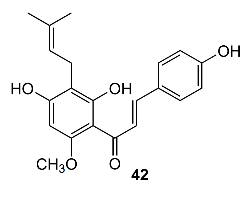
For the study, the authors used a dynamic biofilm model, in which a liquid inoculum (Bacterial inoculum supplemented with curcumin and xanthohumol in BHI liquid nutrient medium (heart and brain infusion agar).) is pumped into a bioreactor (This dynamic in vitro biofilm model consists of a sterile vessel in which bacterial inoculum is placed in a modified BHI liquid nutrient medium. This medium is then pumped by a peristaltic pump at constant pressure into a bioreactor (Lambda Minifor© bioreactor) that maintains the nutrient medium at 37 °C and pH = 7.2 and constant anaerobic conditions (constant flow of a gas mixture of 10% H2, 10% CO2, and 80% N2), thus mimicking the oral environment. This nutrient medium is then continuously transferred (30 ml h–1) via another peristaltic pump to two Robbins devices placed in series in a laboratory oven to maintain a controlled temperature (37 ºC) and containing sterile implants under anaerobic conditions.) under anaerobic conditions maintained by a constant flow of a gas mixture (10% H2, 10% CO2, and 80% N2), and after culturing, microorganisms are fed into a Robbins device carrying an implant. However, the dynamic model of biofilm in vitro proposed by the authors does not fully reproduce the conditions of the human oral cavity. In addition, it is necessary to study the mechanism of biological activity of curcumin (2) and xanthohumol (42) and their effect on the colour and aesthetic quality of implants.
To use dentifrices containing plant extracts, in addition to the assessment of antifungal effect, it is necessary to assess the anti-inflammatory properties in relation to the soft tissues of the prosthetic bed and the ability to prevent bad breath. In turn, for the use of modified acrylic compositions as dentures, in addition to microhardness and flexural strength, their water absorption, water solubility, colour fastness, crack resistance, elastic modulus, and residual monomer content, which affects allergenicity and toxicity, must be studied. Currently, there is no model in implantology that can fully simulate the fluid dynamics and environmental conditions of the human oral cavity.
8. Conclusion
Dental filling materials containing plant extracts of Dioscorea altissima, Olea europaea, Ficus carica, Salvadora persica, Ziziphus spina-christi and Triphala have a strong antibacterial effect against cariogenic S. mutans bacteria, which makes them promising for the use in restorative dentistry. However, dental filling materials themselves may have antibacterial effects, for example, due to the release of some ions and the presence of active functional groups, residual monomers and other unreacted substances, etc.; therefore, it is necessary to know their contribution to the overall antibacterial effect. For this area of dentistry, it is important to conduct clinical studies of new materials, assessing the effect of additives on the curing rate, physicomechanical properties (for example, Vickers hardness).
In the field of endodontics, plant extracts of Thymus lamiaceae, Origanum vulgare, Psidium guajava L., Moringa oleifera, Epilobium parviflorum Schreb, Lawsonia inermis and Boehmeria macrophylla are promising as irrigants. Despite the well-studied antibacterial effect of plant extract-based irrigants on biofilms, their use in endodontics requires randomized clinical trials, studies of their toxicity to human oral mucosa fibroblasts, toxicological tests on animals, and studies of the interaction of irrigants with root canal dentin.
In the field of therapeutic dentistry, extracts of Quercus infectoria, Scrophularia striata, Clitoria ternatea, Zanthoxylum achantopodium, Sambucus williamsii var. Coreana and Lespedeza cuneata are promising for the prevention and treatment of caries, periodontal diseases and wound healing. In this area of dentistry, unlike other areas, clinical randomized studies involving patients with gingivitis have been performed. However, it is necessary to investigate possible adverse reactions over a long period of observation, such as allergic reactions.
In the field of orthodontics, extracts of Cinnamomum verum, Azadirachta indica, toothpastes with the addition of Melaleuca alternifolia, herbal formulas Padma hepaten and Triphala have proved to be effective. In this area, studies of the effect of extracts on the physicomechanical (flexural strength) and adhesive properties of orthodontic appliances and adhesives have been reported, but there is no data on the type of destruction of the adhesive joint, or studies of the effect of additives on the curing rate of acrylic resins.
In the field of dental prosthetics, extracts of Turbinaria conoides, Cinnamomum zeylanicum and Chamaecyparis obtusa can be used. However, in this area of dentistry, additional study of the effect of additives on the curing parameters of acrylic resins (curing rate, curing time, etc.), sanitary and chemical studies in relation to artificial saliva and plasma are required, since residual monomers and secondary metabolites can enter the human body through saliva.
We can conclude that the prospects for using natural additives based on plant extracts and propolis are quite broad. However, to introduce these additives into clinical practice, fairly extensive additional research is required regarding the mechanism of action of active metabolites and their effect on the properties of the final material. Namely, toxicological studies are needed, studies of the allergic and irritant effects of natural additives on tooth tissue, studies of their effect on the mechanical and chemical characteristics of dentin, and in vivo studies with a large sample size. In addition, since dental infections are caused by biofilms rather than single microorganisms, future research should focus on biofilm-based models.
It also seems promising to develop computer models to predict the effect of a drug based on a single natural component on the antimicrobial activity and, accordingly, the possibility of using this drug in a particular area of dentistry and in other fields. Knowing the composition of extracts of particular plants and the properties of these components, it will be possible to optimize the formulation of therapeutic agents, to use only the necessary natural components and/or their mixtures, and to determine which additional components can and/or should be introduced into a product to improve its properties.
9. List of abbreviations
CFU — colony forming units,
DNA — deoxyribonucleic acid,
GIC — glass-ionomer cement,
MIC — minimum inhibitory concentration,
PMMA — poly(methyl methacrylate),
ROS — reactive oxygen species.
Acknowledgements
The authors express their gratitude to the Center of Collective Use of Mendeleev University of Chemical Technology (the State contract 13.CCU.21.0009).
References


Antimicrobial potential of the extracts of the twigs of Azadirachta indica (Neem): an in vitro study