Keywords
Abstract
Nanomaterial-based biosensors have advanced rapidly as transformative technology in clinical diagnostics, offering unparalleled sensitivity, selectivity and versatility. This review provides a comprehensive analysis of the latest developments in biosensors incorporating nanostructured materials, with a focus on their clinical applications. We discuss the wide variety of nanomaterials used in biosensor production, such as carbon-based nanomaterials, metal nanoparticles, quantum dots and two-dimensional materials like graphene and transition metal dichalcogenides. Furthermore, we examine how these nanomaterials are integrated into different biosensing platforms, including electrochemical, optical, and surface plasmon resonance sensors. We emphasize their ability to rapidly and accurately detect clinically relevant biomarkers and analytes. The review offers an in-depth evaluation of the current state of nanostructured biosensor technology and pinpoints critical areas for future research and innovation in this rapidly evolving field.
The bibliography includes 275 references.
1. Introduction
Biosensors offer high specificity and sensitivity in the detection of biological compounds, using physicochemical transducers to convert complex bioanalytical measurements into a user-friendly format. Consequently, biosensor technology has been widely adopted for use in various applications, including diagnostics, environmental monitoring, healthcare, food safety, security and defense.[1] Biosensors provide alternative solutions to address the limitations of conventional detection methods in clinical applications, offering advantages such as high sensitivity, multiplexed detection, rapid response times, low cost, flexibility for on-site testing and continuous monitoring.[2]
A biosensor typically consists of four main components:
(a) a bioreceptor, such as antibodies, nucleic acids, enzymes, aptamers, nanoparticles or cells, with a strong binding affinity for the target analyte;
(b) a transducer, which converts the detected signal into a useful output;
(c) an amplifier, which amplifies and processes the signal;
(d) a display component, which presents the output signal in predefined formats, such as visual, graphical or numeric (Fig. 1).[3][4] The recent integration of nanomaterials has transformed the performance of biosensors, particularly in clinical diagnostics. Spanning zero to three dimensions, nanomaterials offer high surface-to-volume ratios, robustness, enhanced conductivity and tunable optical properties, making them valuable in biosensing applications.[5][6] Advancements in biosensor technology that substantially enhance sensitivity, multiplex detection, real-time monitoring, cost-effectiveness or portability are revolutionizing the field. Recent breakthroughs include ultra-sensitive biosensors capable of detecting trace amounts of biomarkers, miniaturized lab-on-a-chip platforms and wearable biosensors for continuous health monitoring. These advancements have expanded the range of applications for biosensors beyond traditional laboratory settings, enabling rapid diagnostics at the point of care.
![[{"id":"tT1chNzgEA","type":"paragraph","data":{"text":"Principle composition of biosensors"}}]](/storage/images/resized/BpOz1XhMr2BmyD13fowhSUmKYzOvPJqqWtvJg2TB_xl.webp)
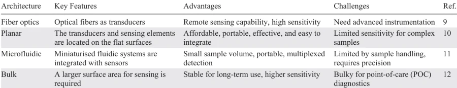
The performance of a biosensor is also strongly influenced by its architectural design. The most common architectures include fiber-optic, bulk, planar and microfluidic systems, each of which has unique advantages and limitations depending on the sensing application.[7][8] Table 1[9-12] summarizes the different biosensor architectures, and highlights their key characteristics, advantages, and challenges.
Nanomaterials, including carbon-based materials, metal nanoparticles (such as silver, gold and platinum) and quantum dots, are often used in the development of biosensors due to their stability, large surface area, high sensitivity to detection and superior electrical and optical properties.[13-19] Although these nanomaterials have been studied for decades, recent advances in their synthesis, surface functionalization and hybrid structure have improved the performance of biosensors. Modifications such as the immobilization of biomolecules on surfaces, doping and the formation of nanocomposites have enabled lower detection limits, increased selectivity and faster response times. Not only do nanomaterials serve as structural components, they also act as transduction elements, enhancing the efficacy of signal conversion. Furthermore, their extensive surface area facilitates biomolecule immobilization and desired electrochemical reactions.[20-22]
Despite their potential, there are challenges associated with integrating nanomaterials into biosensors, including reproducibility and consistency in nanomaterial synthesis, scalability, and mitigating matrix interference. However, recent advancements such as automated synthesis methods, hybrid nanomaterial development and surface functionalization strategies have demonstrated their ability to overcome these limitations. Furthermore, cutting-edge solutions, such as the development of point-of-care (POC) devices and the integration of artificial intelligence (AI)-driven data analysis platforms, are expected to enhance the applicability and effectiveness of nanomaterial-based biosensors in clinical settings.
Although several comprehensive reviews have explored the role of nanomaterials in biosensors for clinical diagnostics,[23-28] these mostly focus on specific concerns. By contrast, this review takes a broader approach, analyzing the application of various nanomaterials in different biosensing platforms and highlighting their suitability for diverse clinical applications. Furthermore, this review explores emerging fabrication methods and the integration of smartphone platforms to enhance the portability and performance of biosensors. We also discuss surface modification strategies to address issues such as non-specific binding and matrix interference, which are critical for clinical applications. By providing a thorough overview of nanomaterial-based biosensors, this review seeks to bridge the gap between fundamental science and practical diagnostic applications. It offers valuable insights into potential future research directions for developing next-generation biosensors.
Unlike earlier studies, which focused primarily on the fundamental properties of nanomaterials, recent research has demonstrated their potential in highly specific biosensor configurations. These include bio-functionalized nanostructures, signal amplification strategies and hybrid nanomaterials with superior transduction efficiency. This review evaluates the outcomes of recent studies on nanomaterial-based biosensors for clinical diagnostics. In this review, we define ‘recent innovations’ as developments that have emerged within the last 5 – 10 years. These innovations have led to significant advancements in nanomaterial synthesis, surface functionalization and the development of hybrid structures that are tailored for use in clinical diagnostics. The review begins with an overview of the different types of nanomaterial, followed by a detailed examination of their applications in biosensing. We then discuss how these nanomaterials interact with analytes, either directly or via functional groups attached to their surfaces. Finally, we highlight their potential clinical applications, address existing challenges and propose future research directions for developing next-generation biosensors that incorporate nanomaterials.
2. Nanomaterials for biosensor fabrication
2.1. Carbon-based nanomaterials
Carbon-based nanomaterials, including graphene, single-walled carbon nanotubes (SWCNTs), multi-walled carbon nanotubes (MWCNTs), fullerenes, carbon nanodiamonds (CNDs), carbon dots (CDs), and graphene quantum dots (GQDs),[29-35] have been extensively studied for use in biosensor fabrication thanks to their unique physicochemical properties and nanoscale dimensions. They possess a high surface area, mechanical strength, superior electrical conductivity and good biocompatibility, making them potential candidates for enhancing the efficacy of biosensors.
Thanks to its two-dimensional structure and high electrical conductivity, graphene facilitates efficient electron transfer and provides ample functional sites for binding analytes. Covalent modifications and π – π interactions further improve graphene’s functionalization potential by enabling the binding of analytes and enhancing sensor efficacy. The π – π stacking interaction between hemin and graphene oxide has been utilized for the simultaneous detection of ascorbic acid, dopamine and uric acid, with limit of detection (LOD) values of 0.3, 0.17 and 0.17 mM respectively.[36] Similarly, graphene and carbon QDs exhibit excellent photoluminescent properties, enabling their use in electrochemiluminescence and fluorescence-based biosensors.
Due to their high aspect ratios and electron transport properties, SWCNTs and MWCNTs contribute to enhancing electrochemical activity and boosting the electron transfer rate of targeted analytes.[37] Carboxylated-MWCNTs coated on indium-tin oxide (ITO) have been used to immobilize aflatoxin B1 (AFB1) antibodies, thereby increasing the stability and sensitivity of the sensor for AFB1 detection with a LOD of 0.08 ng mL–1.[38] Similarly, MWCNTs modified with aflatoxin oxidase on the surface of a Pt electrode resulted in a low LOD of 1.6 nM and a response time of 44 s, demonstrating enhanced sensitivity and rapid analytical performance. The covalent linkage between the MWCNTs and the aflatoxin oxidase improves the enzyme’s activity and facilitates the oxidation of AFB1.
The functionalization of carbon-based materials boosts their physicochemical properties and presents additional active sites for analyte attachment. Covalent and non-covalent functionalization with heteroatoms (such as B, S, and N), biopolymers, metal nanoparticles (NPs), and metal oxide nanoparticles significantly improves the specificity, sensitivity, and stability. For example, MWCNTs combined with conducting polymers such as poly(5,2'-5',2''-terthiophene-3'-carboxylic acid) (pTTCA) have been used to develop a lactate biosensor, in which the covalent bonding between pTTCA and lactate dehydrogenase enhances electron transfer, and achieves a LOD of 1.0 mM.[39]
Moreover, incorporating biopolymers such as polyvinyl alcohol (PVA), polyethylene glycol (PEG), chitosan or poly(3,4-ethylenedioxythiophene): polystyrene sulfonate (PEDOT:PSS) improves the chemical stability and dispersion of graphene oxide (GO) nanoparticles in physiological solutions.[40][41] Polypyrrole (PPy)-based conducting polymers doped with lactate anions demonstrate high selectivity for the detection of lactic acid in human sweat, making them well-suited to non-enzymatic approaches. However, non-enzymatic approaches can introduce challenges relating to the oxidation of other metabolites in complex biological matrices.[42] To overcome this limitation, the incorporation of metal oxide nanoparticles, such as ZnO NPs with MWCNTs, has been shown to further improve sensitivity for lactate detection up to 4.0 nM via electrochemiluminescence.[43]
Integrating carbon-based nanomaterials with biosensing technology enables them to be used in a variety of analytical methods, thereby enhancing detection limits and sensitivity. Electrochemical impedance spectroscopy (EIS), differential pulse voltammetry (DPV) and cyclic voltammetry (CV) are widely used to evaluate the performance of carbon-based biosensors. The remarkable electron transfer abilities of CNTs and graphene improve the sensitivity and response times of electrochemical sensing platforms. Additionally, GQDs and CDs offer real-time and label-free detection of target analytes due to their photoluminescence and fluorescence properties and are commonly used in optical sensing. Furthermore, to ensure the optimum sensor performance, surface morphology and functionalization efficiency are often analyzed by employing transmission electron microscopy (TEM), scanning electron microscopy (SEM), X-ray photoelectron spectroscopy (XPS), and atomic force microscopy (AFM).
Carbon-based biosensors have demonstrated remarkable specificity and sensitivity when detecting various analytes, including pharmaceutical residues, pathogens (such as viruses and bacteria), hazardous ions (e.g. heavy metal ions) and clinical biomarkers.[44-54] Graphene-based field-effect transient (GFET) biosensors have been developed to detect SARS-CoV-2 in human nasopharyngeal swab samples. These biosensors have achieved a LOD of 100 fg mL–1 and 1.0 fg mL–1 in clinical samples and transport medium, respectively.[55] Fig. 2 illustrates the operational procedure of the biosensor. SARS-CoV-2 antibodies were immobilized onto the device using 1-pyrenebutyric acid succinimide ester, an efficient interface coupling agent.
![[{"id":"sHFuQ2cpkZ","type":"paragraph","data":{"text":"Schematic illustration of graphene-based FET biosensor for SARS-CoV-2 detection. Reproduced with permission from the Ref. 55"}}]](/storage/images/resized/UeavcKwMCZENuSivDI1blcN2L8jRuY0hErG9Al9r_xl.webp)
Furthermore, the incorporation of AuNPs into graphene-based FET biosensors has improved the selectivity and sensitivity with which streptavidin and complementary DNA hybridization can be detected, achieving a LOD of 15 aM.[56] The AuNPs facilitate better biomolecular interactions and increase the active surface area, enabling faster electron transfer rates and improving sensor performance.
Integrating carbon-based nanomaterials with biosensing technologies has substantially advanced the field of biosensors, enabling the highly selective, sensitive and multiplexed detection of multiple substances. Their high surface area, excellent electron transfer characteristics and ease of functionalization provide a robust platform for developing real-time and POC applications. Coupling these nanomaterials with analytical methods improves sensor efficiency further by ensuring high stability and sensitivity. Optimizing the integration of carbon-based nanomaterials with evolving sensing platforms and discovering novel functionalization strategies will further enhance the capabilities of biosensors, paving the way for next-generation environmental and clinical monitoring technologies. Furthermore, these materials’ adaptability and flexibility permit the development of portable, miniaturized biosensors, making them ideal for real-time diagnosis in settings with limited resources.
2.2. Metal nanoparticles
In recent years, the term ‘metal nanoparticles’ has emerged in the field of nanotechnology. These particles are made from noble metals that have positive health effects, such as platinum, silver and gold. Researchers have focused on synthesizing metal nanoparticles due to their clear benefits for sensor technology, disease diagnosis and treatment. This section discusses these metal nanoparticles and their relevance to biosensing applications.
2.2.1. Gold nanoparticles (AuNPs)
Gold nanoparticles (AuNPs) have significant potential for use in the design of biosensors thanks to their unique physicochemical properties. Their high electrical conductivity, high surface-to-volume ratio and exceptional surface plasmon resonance (SPR) phenomena make them ideal for enhancing biosensor performance.[57][58] The localized surface plasmon resonance (LSPR) exhibited by AuNPs allows them to induce a characteristic color change (from red to blue) as they transition from a monodispersed to an aggregated state, providing a visual cue for the detection of analytes.[59][60] AuNPs exhibit both LSPR and SPR phenomena, enabling them to produce surface plasmons without external assistance and offering advantages in both electrochemical and optical sensing.[61] The LSPR performance of AuNPs can be tuned by controlling their shape, size and surface modifications, making them highly responsive to changes in the local refractive index. These characteristics are widely used in SPR-based biosensors to detect antibody-antigen interactions, DNA hybridization and small molecules.
AuNPs exhibit diverse morphologies, such as nanorods, nanospheres, nanobipyramids and nanostars. This morphological diversity significantly affects the LSPR phenomenon. This structural diversity enables selectivity and sensitivity to be tailored in biosensing applications.[62][63] For example, SPR-based biosensors use the LSPR of AuNPs to measure analytes by monitoring changes in the refractive index at the sensor surface. Optical fibre SPR biosensors using AuNPs have demonstrated high sensitivity, with a LOD of 1.0 pM for DNA hybridisation,[64] making them ideal for the label-free, highly sensitive and real-time detection of biomolecules.
The surface modification of AuNPs with biomolecules such as aptamers, DNA probes, enzymes and antibodies enhances their biorecognition capability.[65] Covalent and non-covalent interactions, including hydrophobic and electrostatic forces, can increase the sensitivity and specificity of sensors. For instance, aptamer-functionalized AuNPs have been used to diagnose various biomarkers, including cancer biomarkers with LOD as low as 1.0 pg mL–1, and microRNAs (miRNAs) with remarkable LODs down to 100 aM,[66][67] respectively. AuNPs can bind to biomolecules due to their strong affinity for thiols, amines, and disulfides. Consequently, AuNPs can trap a large number of biomolecules while retaining bioactivity on biosensor surfaces. Self-assembled monolayers of thiol-containing biomolecules on the surface of AuNPs facilitate stable and robust functionalization, ensuring reproducible and reliable sensing.
AuNPs exhibit a visible color change when they interact with or aggregate around target substances, a property that has been widely exploited in colorimetric biosensors. This phenomenon has been used to measure pathogens, DNA hybridization and biomarkers. For instance, Behrouzi and Lin designed a colorimetric plasmonic biosensor using AuNPs to detect SARS-CoV-2 nucleocapsid protein, achieving an LOD of 150 ng mL–1 in five min.[68] Similarly, AuNPs have been employed to detect cancer biomarkers (CD44) with a LOD of 0.11 pM.[69]
AuNP-Modified electrodes increase electron transfer rates and enable highly sensitive electrochemical detection. Techniques such as EIS, CV and DPV are commonly used to analyze target substances with the aid of AuNPs. AuNP-Modified carbon screen-printed electrodes (SPEs) have successfully detected Escherichia coli O157:H7 strain in concentrations ranging from 10 to 106 colony-forming units (CFU) per mL, with a LOD of 15 CFU mL–1 and a detection time of approximately 30 min.[70]
AuNPs conjugated with luminophores or fluorophores are used in electrochemiluminescence (ECL) and fluorescence-based biosensors to enable the sensitive analysis of target molecules. Combining AuNPs with QDs enhances their photoluminescence properties, making them ideal for the multiplexed detection of various molecules.[71-74] For instance, a hybrid system of GO, GQDs, and AuNPs was used to detect microRNAs (miRNAs-210, miRNAs-21, and miRNAs-155) for breast cancer diagnosis, resulting in high sensitivity (Fig. 3).[74] Firstly, the thiol-modified miRNA probes for the three target miRNAs were treated with tris(2-carboxyethyl) phosphine hydrochloride to reduce the disulfide bonds and activate the probes for immobilization. The surface of the screen-printed carbon electrode (SPCE) was then modified using a combination of anthraquinone (AQ), methylene blue (MB) and polydopamine (PDA) as redox species, as well as AuNPs, GO and GQDs. Reduced thiol-modified capture probes that were specific to miRNAs 210, 21 and 155 were then immobilised onto the modified SPCE surface in order to develop a three-miRNA probe-modified array. Square wave voltammetry (SWV) was used to record the response and assess the electrochemical performance of the biosensor. The developed biosensor exhibited a wide concentration range of 0.001 – 1000 pM, with notable LODs of 0.28 fM, 0.04 fM and 0.33 fM for miRNA-210, miRNA-21 and miRNA-155, respectively.
![[{"id":"xKnKcvgjXi","type":"paragraph","data":{"text":"AuNPs/GO/GQDs-based biosensors for detecting miRNA-155, miRNA-210, and miRNA-21 breast cancer biomarker. Reproduced with permission from the Ref. 74. The electrochemical cell consisted of a working electrode (WE), counter electrode (CE), and reference electrode (RE). 6-Mercapto-1-hexanol (MCH) was used as a blocking agent to prevent nonspecific binding on the sensor surface."}}]](/storage/images/resized/xkU2BDfQ7QpkkTY6PPJwlwH2NvJl7nQy1hlLbvmB_xl.webp)
A biosensor based on PPy/graphene particles (GP)/AuNPs was successfully developed for the detection of miRNA-21, achieving an impressive LOD of 0.020 fM and a linear range from 1.0 fM to 1.0 nM (Fig. 4).[75] In this design, a SPCE was modified with a GP/PPy nanocomposite to enhance electron transfer and facilitate uniform dispersion of AuNPs. Capture DNA-21 probes (complementary to the target miRNA-21) were immobilized on the electrode surface along with AuNPs, followed by blocking with MCH to prevent nonspecific adsorption. After hybridization with the target miRNA-21, MB was intercalated, and the final DPV signal was recorded. The peak current from the MB redox process, which correlates with the degree of hybridization, served as the quantitative signal for miRNA-21 detection. AuNP-based biosensors have demonstrated excellent sensitivity for COVID-19-related biomarker detection. For instance, a dual-mode biosensor utilizing both colorimetric and electrochemical approaches enabled rapid SARS-CoV-2 antigen detection, achieving limits of detection (LODs) of 48 ng mL–1 (colorimetric) and 1.0 pg mL–1 (electrochemical), respectively.[76] Similarly, AuNPs coupled with nitrogen-doped carbon dots (N-doped CDs) were employed for the detection of interleukin-6 (IL-6), a key inflammatory biomarker associated with SARS-CoV-2 infection, attaining a LOD of 0.82 pg mL–1.[77]
![[{"id":"Wy6bzjacg9","type":"paragraph","data":{"text":"Schematic steps of biosensor fabrication and miRNA detection: (<i>1</i>) modification of the GP/PPy composite on a surface of the SPCE; (<i>2</i>) deposition of adsorbed ss-DNA-21 probe/AuNPs on a modified electrode; (<i>3</i>) adding MCH blocking solution; (<i>4</i>) adsorption of MB into capture DNA-21/AuNPs/GP/PPy/SPCE; (<i>5</i>) capturing the miRNA-21 target at the DNA-21 probe; (<i>6</i>) adsorption of MB into miRNA-21/capture DNA-21/AuNPs/GP/PPy/SPCE; (<i>7</i>) recording DPV responses of the electrodes before and after hybridization. Reproduced with permission from the Ref. 75"}}]](/storage/images/resized/dGTrxpluwNsJ3xG3pO7oBx8jsL87ovJUrjgboCO6_xl.webp)
Au-based nanostructures significantly enhance SERS by generating plasmonic hotspots, thereby enabling highly sensitive and specific detection for early disease diagnosis and POC applications. For instance, Au-semi-coated polystyrene nanospheres were used to optimize photonic crystal configurations, resulting in a 790-fold enhancement of the Raman signal with high stability.[78] This platform has been applied to melanoma diagnosis, achieving two to three orders of magnitude greater sensitivity than conventional SERS substrates. Furthermore, a smartphone-integrated SERS biosensor was recently developed for the simultaneous detection of Escherichia coli O157:H7, Pseudomonas aeruginosa, and Staphylococcus aureus by leveraging CRISPR/dCas9*[79]-guided hotspot self-assembly. In this context, AuNPs amplify the SERS signal by creating dense hotspots, which facilitate multiplexed detection with high specificity.
Future research should focus on improving the selectivity and sensitivity of AuNP-based biosensors further by exploring novel surface modification strategies that can modulate the LSPR behavior of AuNPs and increase their functionalization efficiency. Various AuNP morphologies should be studied to tailor the efficacy of biosensors for specific applications. Another area of focus should be developing hybrid sensing platforms that combine AuNPs with advanced materials such as MXene and conducting polymers to attain enhanced performance. The miniaturization of biosensor platforms continues to enhance their applicability for POC and real-time applications in environmental and clinical monitoring. It is notable that many AuNP-based lateral flow immunoassay techniques are already widely implemented in POC diagnostics, underlining their established role in current healthcare and field-testing scenarios.[80]
* CRISPR/dCas9 is a catalytically inactive variant of the CRISPR associated protein Cas9, which can bind to specific DNA sequences without cutting them, enabling programmable biosensing application.
2.2.2. Silver nanoparticles (AgNPs)
Silver nanoparticles (AgNPs) have also attracted considerable interest in the fabrication of biosensors due to their unique physicochemical properties, including high catalytic activity, electrical conductivity and LSPR. These properties enhance the selectivity, sensitivity and analytical efficacy of biosensors. Compared to other noble metals, such as Au, AgNPs exhibit narrower plasmonic bands, superior plasmonic effects, and a higher refractive index sensitivity, making them ideal for plasmonic biosensors.[81] The LSPR properties of AgNPs can be precisely tuned by controlling their shape, size, and the dielectric environment in which they are embedded, all of which play an important role in developing highly sensitive plasmonic biosensors. Morphologies such as nanorods, nanoflowers and nanoprisms exhibit distinct plasmonic responses that can be optimized for the detection of specific targets. Zhou et al.[82] demonstrated this potential by designing an AgNP array-based LSPR biosensor in the form of a triangular structure for detecting serum p53 protein in head and neck squamous cell carcinoma. Using this biosensor, they showed a significant LSPR shift between cancer and healthy patient samples. This was one of the earliest clinical applications of LSPR biosensors in cancer diagnostics, validating the role of AgNPs in sensitive biomarker detection. Another approach involves using stimuli-responsive hydrogel-AgNP nanocomposites in LSPR-based optical biosensors to enhance the sensitivity and selectivity of glucose detection. These hydrogels swell upon analyte recognition, thereby modulating LSPR absorbance and improving the detection limits.[83]
AgNPs exhibit remarkable catalytic and electrical properties that accelerate electron transfer kinetics in electrochemical biosensors. These features enhance signal transduction and improve detection limits by minimizing electron transfer resistance. Furthermore, combining AgNPs with graphene, MWCNTs and polydopamine can enhance their electrochemical properties by improving conductivity and increasing the surface area. Wang et al.[84] designed a non-enzymatic glucose biosensor by integrating Pt and reduced graphene oxide (rGO) with AgNPs, achieving a sensitivity of 129.32 μA mM–1 cm–2 and a LOD of 1.8 mM, demonstrating the synergistic effects of these materials. Similarly, AgNPs were adsorbed directly onto MoS2 micro flowers on a Pt electrode for non-enzymatic glucose detection, achieving an LOD of 1.0 mM,[85] which emphasizes the clinical potential of AgNPs-based nanocomposites.
The surface modification of AgNPs plays a dynamic role in enhancing selectivity and stability and facilitating the binding of biorecognition elements. For example, a graphene-polydopamine (PDA)-AgNPs composite was developed for the selective and sensitive detection of DNA.[86] Fig. 5a illustrates the step-by-step fabrication process. The AgNPs-PDA/graphene (noted as Ag-pdop@Gr in Fig. 5a) solution was drop-coated onto a glassy carbon electrode (GCE). Thiolated single-stranded DNA (HS-ssDNA) probes were immobilized on the electrode surface via Au-thiol bonding to form ssDNA-S-AgNPs-PDA/graphene. To reduce nonspecific adsorption, the surface was treated with MCH. Hybridization was then performed in phosphate buffer with complementary target DNA (cDNA), and the unbound sequences were removed by washing. MB was used as an electrochemical indicator, achieving an LOD of 3.2 × 10⁻¹⁵ M. This biosensor also demonstrated excellent selectivity by distinguishing even one-base mismatched sequences, highlighting its potential for nucleic acid detection. Similarly, a glucose biosensor was designed by immobilizing glucose oxidase (GOx) on AgNPs decorated MWCTs modified GCE. The AgNP-MWCNT composite membrane improved biocompatibility for GOx immobilization and enhanced electrocatalytic activity toward oxygen reduction. Notably, the AgNPs facilitated direct electron transfer between the redox-active site of GOx and the GCE surface due to their excellent conductivity and high protein-loading capacity, resulting in direct electrochemistry of GOx. This biosensor demonstrated a low LOD of 0.01 mM and a wide linear range from 0.025 to 1.0 mM, with good stability and reproducibility.[87] Recently, a multifunctional hydrogel sensor was fabricated by incorporating Ag-loaded PDA NPs (Ag@PDA) into a thermally cross-linked acrylamide – methacrylamide – chitosan (CSMA) and polyacrylamide (PAM) network, referred to as Ag@PDA/(CSMA-PAM) (Fig. 5b).[88] This hydrogel exhibited high sensitivity to both mechanical stimuli: a pressure sensitivity of 0.07 kPa–1 in the range of 0 – 2.15 kPa, and a strain sensitivity strain of 2.13 over the range of 65 – 150%, indicating reliable responsiveness to large stretching deformations. It also showed fast response and recovery times (136 ms for pressure, 550 ms for strain). Furthermore, the presence of Ag@PDA endowed the hydrogel with biocompatibility, antibacterial, and antioxidant properties, making it highly suitable for wearable sensor applications in personalized healthcare.
![[{"id":"iXR1_LN_J1","type":"paragraph","data":{"text":" (<i>a</i>) Schematic illustration of the electrochemical DNA biosensor using Ag-PDA / Graphene. Reproduced with permission from the Ref. 86. (<i>b</i>) Graphic diagram of the Ag@PDA/(CSMA-PAM hydrogel as multifunctional pressure/strain sensor. Reproduced with permission from the Ref. 88."}}]](/storage/images/resized/PUghkORka1zLmBFPlgFajDCEIm4rGtomezIydLbZ_xl.webp)
AgNPs play a crucial role in the design of label-free SERS sensors, substantially increasing their sensitivity and broadening their range of applications. One notable example is the use of bimetallic Au-Ag nanocuboids, which made it possible to fabricate a highly sensitive label-free SERS sensor for the ultrasensitive detection of florfenicol residues in eggs.[89] Furthermore, a ZIP-8/Ag-based SERS sensor demonstrated high sensitivity, detecting urea at concentrations as low as 0.148 nM in standard solutions and 10–8 M in milk.[90] Depending on the target analyte, AgNPs undergo various surface modifications to enhance biocompatibility, minimize fouling and increase stability, thereby validating their potential for clinical diagnostics.
2.2.3. Platinum nanoparticles (PtNPs)
Platinum nanoparticles (PtNPs) are widely used in biosensor applications due to their high electrical conductivity and catalytic efficiency compared to other metal nanoparticles, enabling enhanced electron transfer and improved detection sensitivity. Their high surface-to-volume ratio enhances catalytic efficiency and facilitates faster electron transfer, supporting effective sensor performance. These attributes make PtNPs suitable for detecting a wide range of environmental and biological molecules, including hydrogen peroxide, hydrazine, ascorbic acid and glucose. To maximize these benefits, PtNPs are often dispersed onto cost-effective substrates such as MWCNTs, GO, and polymeric composites, which help to reduce costs, optimize Pt usage, maintain catalytic activity, and prevent NPs aggregation.[91-96]
The catalytic efficiency of PtNPs can be further tailored by modifying their shape, size, and surface chemistry. Furthermore, combining PtNPs with other materials, such as AuNPs, graphene and conducting polymers, can result in synergistic effects that enhance stability, conductivity and electron transfer rates, thereby improving analytical performance. In particular, carbon-based materials serve as excellent support matrices for PtNPs by preventing aggregation and improving their electrochemical properties, offering advantages over other supports such as metal oxides and polymers due to their superior conductivity and surface area.[97]
In electrochemical biosensors, PtNPs help to reduce overpotentials and accelerate electron transfer, thereby increasing signal transduction efficiency. Electrochemical methods such as EIS, CV and DPV are commonly used to evaluate the analytical performance of PtNP-based biosensors. In addition to electrochemical methods, PtNPs have been used in SPR and LSPR biosensors, where their unique optical properties enable increased refractive index sensitivity and more selective biosensing capabilities.
PtNPs are particularly prevalent in glucose biosensors, where their high catalytic activity towards glucose oxidation results in increased sensitivity. These sensors can operate using either an enzymatic or a non-enzymatic approach. In enzymatic systems, GOx is often immobilized on PtNP-modified electrodes to promote glucose oxidation. For instance, a glucose sensor was developed using PtNPs that were electro-deposited onto poly(Azure A), followed by the immobilization of GOx onto activated carbon electrodes. This sensor exhibited good selectivity in real samples, such as plant cell culture medium and commercial juices.[98] Similarly, PtNPs – polyaniline (PANI) – montmorillonite (MMT) nanocomposites and GOx have been shown to enhance electron transfer and electrocatalytic activity, achieving a LOD of 0.1 μM and a range of 10 μM to 1.94 mM.[99] In non-enzymatic approaches, PtNPs can catalyse glucose oxidation directly, without requiring enzymatic activity.[100]
PtNP-Based aptamer biosensors for detecting alpha-fetoprotein (AFP) have been developed (Fig. 6a),[101] using a graphene-carbon paste electrode modified with PtNPs/GO-COOH. This was followed by activation using a mixture of N-ethyl-N'-[3-dimethylaminopropyl] carbodiimide/N-hydroxysuccinimide (EDC/NHS), which enabled aptamer immobilization. NH2-Aptamer immobilization was then followed by blocking of non-specific binding sites with bovine serum albumin (BSA). The resulting label-free biosensor demonstrated a detection range of 3.0 – 30 ng mL–1 and a LOD of 1.22 ng mL–1. It showed high selectivity and accuracy for AFP in human serum samples, making it suitable for the early diagnosis of cancer. The composition of PtNPs and AuNPs has shown great potential in enhancing biosensor performance. Mahobiya et al.[102] exploited this synergy to detect glycated albumin, coating the electrode with a molecularly imprinted polymer (MIP) to achieve high sensitivity and selectivity over a wide concentration range (500 mM to 0.1 nM) with a low LOD of 0.1 nM (Fig. 6b). The micro-screen-printed electrode (μSPE) was modified with Au-PtNPs and electrodeposited with MIP using CV at −0.2 to 0.6 V for 15 cycles. After washing, the imprints improved conductivity, rendering the biosensor effective for glycated albumin detection.[102] Similarly, Hasanjani et al.[103] developed a biosensing probe based on a bimetallic Pt-AuNPs/GO/chitosan/DNA composite that could detect zidovudine at an impressively low level of 0.003 pM. PtNPs have been integrated into multi-analyte biosensors for the simultaneous detection of several biochemical species. Oh et al.[104] developed a sensor using PtNPs and graphene to detect seven biomarkers, including uric acid (UA), ascorbic acid(AA), dopamine(DA), hypoxanthine (HX), nitrite (NO2–) and acetaminophen (AP) in human blood serum (see Fig. 6c). This sensor demonstrated high precision and accuracy, highlighting its potential for environmental and clinical applications. PtNPs/rGO nanocomposites were deposited on the GCE via a one-step electrochemical reduction process to create an electrochemical sensor capable of detecting seven biomarkers. Additionally, the performance of GCEs modified with PtNPs-MWCNTs, AuNPs-MWCNTs and AuNP-PtNPs-MWCNTs for cholesterol detection was investigated.[105] Amperometric biosensors were developed by coating cholesterol oxidase (COx) onto metal/MWCNTs-functionalised electrodes. The COx/Au/MWCNTs electrode demonstrated the most favorable response (see Fig. 6d ), with a LOD of 0.5 mM and a broad linear detection range from 1.4 mM to 2 mM. This enhanced performance is attributed to synergistic effects that improve both electron transfer and sensitivity. Despite their impressive performance, PtNP-based biosensors face challenges relating to cost-effectiveness, reproducibility and long-term stability. Exploring bimetallic materials, optimizing surface modification and hybrid nanostructures, and incorporating miniaturized and flexible platforms could address these limitations. Future research should focus on developing low-cost, scalable fabrication techniques, as well as integrating PtNP-based sensors with smartphone-based platforms for POC diagnostics.
![[{"id":"8xtFykGGrR","type":"paragraph","data":{"text":" (<i>a</i>) A label-free aptasensor for sensitive determination of alpha-fetoprotein based on aptamer-loaded PtNPs/carboxylated-graphene oxide. Reproduced with permission from the Ref. 101. (<i>b</i>) The step-by-step development of PtNPs/AuNPs-based biosensor for determining glycated albumin. Reproduced with permission from the Ref. 102. (<i>c</i>) PtNPs/rGO-based biosensor for the simultaneous detection of seven biochemical substances. Reproduced with permission from the Ref. 104. (<i>d</i>) Calibration curves of the biosensors included COx/Au/MWCNTs electrode. Reproduced with permission from the Ref. 105."}}]](/storage/images/resized/VKXR4QAOgPVR22xQPlv6lSP7WNt1qy2qk3WKhy1p_xl.webp)
2.3. 2D nanomaterials
2.3.1. Graphene
These are graphene-based field-effect transistor (FET)-like structures that employ rGO as sensing elements. These structures have shown promise in detecting metal ions and various other analytes, while also demonstrating the versatility and potential of graphene in biosensing applications.[106-108] rGO is easily prepared and rich in oxygen functional groups, making it useful for immobilizing biomolecules. As the first 2D material to be discovered and utilized, graphene stands out as the only 2D material with considerable potential for large-scale commercial applications to date.[109-111] Despite lacking a band gap, which limits its use in digital circuits, graphene is a popular choice for gate materials in biological field-effect transistors (bioFETs) and POC biomedical devices.
Significant interest in integrating graphene into bioFET technology was sparked by a study by Mohanty et al.[112] using a graphene-based FET (GFET) to detect DNA hybridization.Similarly, a biosensor employing an aptamer-based GFET has been engineered to detect alcohol dehydrogenase (ADH), demonstrating high specificity and sensitivity.[113] The biosensor exhibited a significant increase in current as the concentration of ADH rose from 1 pg mL–1 to 10 ag mL–1, demonstrating a sensitivity of 50.00 mA g–1 mL−1 and a LOD of 3.55 ag mL–1.
GFETs have also been used to detect myoglobin at low concentrations of around 30 fg mL–1, demonstrating high sensitivity and making them suitable for POC cardiac diagnosis and lab-on-chip platforms. In another study, silk protein was used as a substrate and carrier for GOx in the development of a GFET device for glucose detection.[114] This biosensor detects glucose levels by analysing the differential drain-source current and Dirac point shift of the graphene transistor during glucose oxidation by GOx. The LOD of the biosensor was approximately 0.1 mM, recorded at gate voltage (Vg) = 0 V and drain-source voltage (Vds) = 100 mV.
GFET biosensors have also been used to detect heart failure-related biomarkers in whole blood [115] and to perform label-free RNA [116] detection with high selectivity and sensitivity. They can distinguish target RNA from non-complementary RNA, achieving detection limits as low as 0.1 fM, which is two orders of magnitude better than previously reported. Beyond nucleic acids, GFET biosensors have also been employed to recognise target proteins and enzymatic activities such as acetylcholinesterase and inhibition reactions. Another FET-biosensor based on rGO has shown promise as an in situ analytical tool for investigating the impact of drugs on Alzheimer’s disease treatment.[117]
Graphene-based electrochemical sensors are widely used to detect a variety of analytes.[44] [118-120] For example, graphene modified with 1-pyrene butanoic acid succinimidyl ester can selectively detect recombinant cyanovirin-N with a detection range of 0.01 – 10 ng mL–1 and an impressive LOD of 0.45 pg mL–1.[121] Furthermore, porous laser-induced graphene (LIG) incorporating PtNPs, chitosan and the GOx enzyme exhibits high sensitivity (4.622 μA mM–1) and a detection limit of less than 300 nM with a dynamic linear range of up to 2.1 mM for glucose biosensing.[122] Similarly, a chitosan, GOx and LIG graphene electrode on a polyimide substrate achieves a LOD of 0.431 mM for glucose detection.[123] Additionally, graphene-based nanocomposite materials are also being used in wearable smart biosensors for real-time analysis, thus expanding their scope in biosensing.[124]
2.3.2. Transition metal dichalcogenides (TMDs)
Transition-metal dichalcogenides (TMDs), represented by the general formula MX2, where M is a transition metal and X is a chalcogen (Te, Se or S), have attracted significant interest for use in biosensing applications due to their diverse electronic and structural properties.[19][125] These materials exhibit a variety of behaviours, including insulating (HfS2), semiconducting (WS2, MoS2), metallic (VSe2, NbS2), and semimetallic (TiSe2, WTe2) behaviours, which makes them highly adaptable for biosensor fabrication.[126-130]
The unique layered structure of TMDs, consisting of covalently bonded MX2 layers held together by weak van der Waals forces, facilitates easy exfoliation to form 2D nanosheets. These structures have a high surface-to-volume ratio, excellent biocompatibility and tunable electronic and optical properties, making them ideal for use in biosensors. Controlling the thickness, lateral dimensions and defect density of TMD nanosheets precisely can significantly enhance their electrochemical and optical properties.[131]Recent research has also explored the properties of 0D and 1D TMD structures, enabling the fine-tuning of their catalytic activity, charge transport and optical behavior. Modifying the crystal structure, doping, or integrating TMDs with other nanomaterials (e.g. graphene or gold nanoparticles) enables researchers to enhance the sensitivity and selectivity of TMD-based biosensors.[132-139]
Several studies have demonstrated the versatility of TMDs in biosensing applications. For example, Chauhan et al.[140] developed a biosensor for cardiac biomarker troponin I using nanostructured Mo3Se4 combined with rGO, achieving a detection range of 1.0 fg mL–1 to 100 ng mL–1 with a LOD of 1.0 fg mL–1. 2D WS2 nanoflakes combined with AuNPs were employed to fabricate a sensor for human serum albumin, with a LOD of 2 ng mL–1, enabling microalbuminuria screening.[141] Similarly, WS2/AuNPs nanocomposites enhanced the photoelectrochemical properties of WS2, achieving a LOD of 0.5 pg mL–1 for carcinoembryonic antigen detection.[142] MoS2 sheets have demonstrated the ability to distinguish between dopamine and ascorbic acid, as well as showing high sensitivity for glucose detection and miRNA determination.[143][144]
TMDs also have significant potential to enhance the sensitivity of SPR biosensors.[143] For example, the integration of MoS2 nanosheets with an Au-coated optical fiber significantly improved the performance of an SPR biosensor for detecting BSA. In this system, the Au-coated fiber surface was modified with MoS2 nanosheets, followed by the immobilization of anti-BSA antibodies (Fig. 7). The enhancement in SPR sensitivity was attributed to the synergistic effects of MoS2 and Au, with MoS2 contributing additional surface area and facilitating biomolecule immobilization, enabling direct, chemical-free antibody binding via a convenient method of hydrophobic interaction. As a result, the MoS2-modified biosensor achieved a lower LOD (0.29 mg mL–1) compared to the unmodified Au sensor (0.45 mg mL–1), demonstrating the efficacy of TMDs in enhancing biosensor performance.[145]
![[{"id":"5yMx0efEI-","type":"paragraph","data":{"text":"Schematic description of the fabrication process for an optical fibre-based SPR biosensor. Method 1 shows the biosensor without MoS<sub>2</sub>, while Method 2 shows the biosensor with a MoS<sub>2</sub>-modified optical fibre SPR configuration. Reproduced with permission from the Ref. 145."}}]](/storage/images/resized/Gkhypq3Q4goacpZBsE4nE9wlW1BjEvzf4dxiZejG_xl.webp)
A similar strategy was employed to develop a highly sensitive, label-free impedimetric sensor for detecting total T3 in human serum samples. AuNPs and MoS2 were electrochemically deposited on ITO substrates, followed by the formation of a monolayer of dithiobis(succinimidyl propionate) (DSP) to bind T3 antibodies (anti-T3) via amine coupling. This formed Au – MoS2/anti-T3 electrode transducers (Fig. 8a). The sensor achieved linear quantification of T3 in the range of 0.01 – 100 ng mL–1, with a LOD of 2.5 pg mL–1.[146] A MoS2 and graphene nanocomposite has also been successful used to detect dopamine with a LOD of 10 pM and has proven effective in analyzing dopamine in real blood samples.[147] Furthermore, a label-free, reliable immunosensor for AFP detection was developed using a smartphone-integrated system. Exfoliated 2D MoSe2 and 2D WSe2 were used to modify an SPCE, which was connected to a small potentiostat linked to a smartphone. This modified SPCE facilitated antibody immobilization and enabled AFP detection via a sandwich-type immunocomplex with the corresponding aptamer (Fig. 8b). The 2D MoSe2/WSe2 heterojunction increased the reactivity of the SPCE, providing a large surface area and high adsorption capacity. The electrochemical response of MB intercalating with the aptamer produced the detection signal, with a linear range of 1.0 – 50 000 pg mL–1 and a LOD of 0.85 pg mL–1.[148] TMDs have also been used in fluorescence biosensors, particularly for nucleic acid analysis. MoS2 nanoflakes have been shown to exhibit high selectivity for DNA targets,[148] permitting reliable fluorescence-based biosensing. Overall, TMDs are proving to be significant materials for biosensing applications, offering high sensitivity and selectivity, as well as potential applications in various biomedical and environmental analyses.
![[{"id":"-l4UiXIwJZ","type":"paragraph","data":{"text":"(<i>a</i>) A schematic illustration of the preparation of the T<sub>3</sub> receptive interface, showing the step-by-step process of MoS<sub>2</sub>/Au formation, followed by functionalization with a DSP layer on the ITO substrate. Reproduced with permission from the Ref. 146. (<i>b</i>) Development of a 2D MoSe<sub>2</sub>/WSe<sub>2</sub> modified electrode for the detection of AFP. Reproduced with permission from the Ref. 148."}}]](/storage/images/resized/7euMwI8m6IFFWfDzPhBUj6se4zcqsO5aG6nnPnlI_xl.webp)
2.4. Quantum dots
Quantum dots (QDs) are quasi-zero-dimensional semiconductor nanocrystals that have emerged as promising candidates for biosensing applications due to their exceptional optoelectronic properties and quantum confinement effects. The quantum confinement effect, which depends on the Bohr radius and particle size, enables precise tuning of emission wavelengths. The broad absorption spectrum of QDs enables excitation at the wide diapason of wavelengths, while their narrow, size-tunable emission spectra allow for the multiplexed detection of different analytes. QDs typically have a core-shell structure: the core determines the optical properties and the shell enhances the quantum yield and improves photostability.[19] [149][150]Their high fluorescence quantum yield, long fluorescence lifetime and large extinction coefficients make QDs ideal for use in electrochemical, photoelectrochemical and fluorescence biosensors.[151-153]
Furthermore, QDs are much brighter than traditional organic dyes, making them highly desirable for use in fluorescence-based biosensing platforms. Another significant advantage of QDs is their surface modifiability, which enables them to be functionalized with biomolecules such as antibodies, aptamers and enzymes. This flexibility enhances the specificity and selectivity of QD-based biosensors, enabling them to be adapted for a wide range of biomedical and environmental applications.
QDs have been widely utilized in various biosensing platforms due to their excellent sensitivity and selectivity. A ZnS/CdSe QD-based nuclear receptor fluorescence probe (QDs-NRFP)-mediated biosensor has been developed to screen for retinoic acid (RA), an endocrine-disrupting chemical.[150] This biosensor was fabricated via an antigen – antibody immunobinding interaction between CdSe/ZnS QD-labelled anti-GST tag antibodies and the GST tag of the human retinoic acid receptor β ligand-binding domain (GST-hRARβ-LBD). The high quantum yield of the CdSe/ZnS QDs increases the GST-hRARβ-LBD binding activity and improves sensitivity, achieving a LOD of 1.8 ng L–1 within a linear range of 7.5 – 1183.6 ng L–1. The biosensor has been shown to be highly reliable and accurate in detecting RA binding activities in both physiological and wastewater samples.[154] In another study, ZnSe@ZnS QDs and Mn-doped ZnS Ds were used as fluorophores to develop a ratiometric fluorescent assay for the detection of alkaline phosphatase (ALP) using p-nitrophenyl phosphate (PNPP) as the substrate. The overlap between the QD excitation spectra and the PNPP absorption spectra resulted in fluorescence quenching. Following the hydrolysis of PNPP by ALP, p-nitrophenol was produced, causing a red shift in the PNPP absorption band and fluorescence recovery of Mn : ZnS QDs (585 nm), while the overlap between the emission spectra of the ZnSe@ZnS QDs and the absorption spectra of p-nitrophenol resulted in further quenching of the ZnSe@ZnS QDs (405 nm). The ratiometric fluorescent signal (F585/F405) was associated with ALP activity and the LOD was found to be 0.57 U per L within a linear range of 4.0 to 96 U per L with an R² value of 0.9969, indicating an excellent linear correlation between ALP concentration and the fluorescent signal. This approach effectively monitored ALP activity in HepG2 cells and human serum.[155]
rGO-QDs decorated with silk fibroin were used to develop a fluorescence turn-on probe for levodopa detection. The sensor exhibited an increase in fluorescence intensity in the presence of levodopa, with the LOD calculated to be 76.18 nM over a linear concentration range of 0 – 35 mM. The sensor was integrated into a smartphone-based device, where a 365 nm light-emitting diode (LED) was positioned beneath the rear camera to capture fluorescence color changes during sensing. Using this setup, a LOD of 0.29 mM was observed within the same concentration range.[156]
QD-Based fluorescent biosensors are also effective in detecting environmental contaminants. A fluorescent biosensor has been fabricated for sensitive and selective detection of Pb2+ ions, employing a DNAzyme system that activates only in the presence of Pb2+. This nanoprobe is based on nitrogen- and boron-doped carbon dots (BNCDs) and a carboxyl-functionalized terbium metal-organic framework, which acts as a fluorescent tag, and an NH3-GR5 DNAzyme, which acts as a bioreceptor quencher. The fluorescent tag exhibited dual emission peaks in the green and blue regions. In the absence of Pb2+, the fluorescence intensity was quenched due to overlap between the BNCDs@Tb-MOF and the quencher. Upon Pb2+ activation, the catalytic core of the DNAzyme cleaves the quencher-tagged substrate strand, thereby restoring fluorescence. The biosensor achieved a LOD of 0.96 ppb over a concentration range of 2.0 to 1000 nM.[157] Additionally, a CdTe QD-based apta-nano fluorescence biosensor was developed for the detection of methamphetamine in biological fluids. The sensor achieved a detection range of 1.34 × 10⁻¹⁰ to 1.24 × 10⁻⁷ M with a LOD of 40.34 × 10⁻¹² M. It used a ligand-exchange process in which the thioglycolic acid molecules on the QD surface were replaced by a thiolated methamphetamine aptamer. The fluorescence intensity of the QDs increased upon aptamer attachment and decreased upon aptamer-methamphetamine complex formation.[158] β-Cyclodextrin-decorated sulphur QDs were employed for the selective fluorescence recognition of tryptophan enantiomers, achieving a LOD of 2.3 nM.[159]
QD-Based electrochemical biosensors have been designed for clinical applications. One such biosensor, designed for glucose detection, uses PbS colloidal quantum dots (PbS CQDs) and gold nanospheres (AuNSs).[160] The PbS CQDs/AuNSs/GOx mixture is immobilised on the carbon electrode through dip-coating. This biosensor integrates the recognition of specific molecules, signal transduction and signal amplification, converting the glucose enzyme-catalysed reaction into measurable current signals. The biosensor exhibited a linear detection range from 0.1 mM to 10 mM, with a LOD of 1.432 mM. Another sensor, which combined QDs with SWCNTs and aromatic dyes, demonstrated exceptional sensitivity for nucleic acid detection. The LOD was 89 aM for miRNA-155 and 64 aM for miRNA-21, with a detection range of 0 to 1.0 pM.[161]A highly sensitive method for detecting tryptophan in serum samples was also developed using a pencil graphite electrode modified with carbon dots and polypyrrole (PPy), demonstrating superior selectivity.[162] Another electro-chemo-luminescence biosensor using carbon QDs was developed for microRNA analysis and showed a LOD of 1.95 fM, highlighting its potential for clinical biomarker detection.[163]
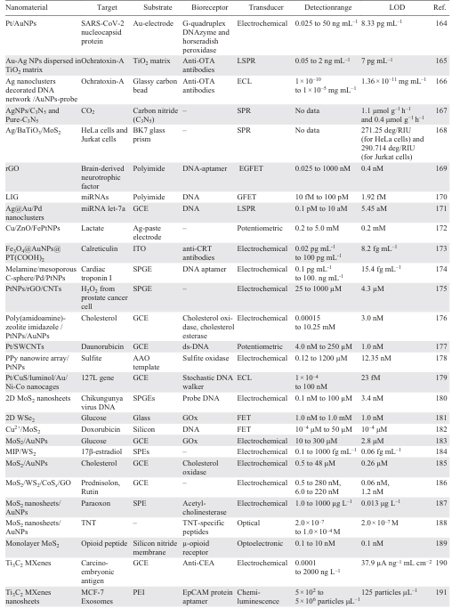
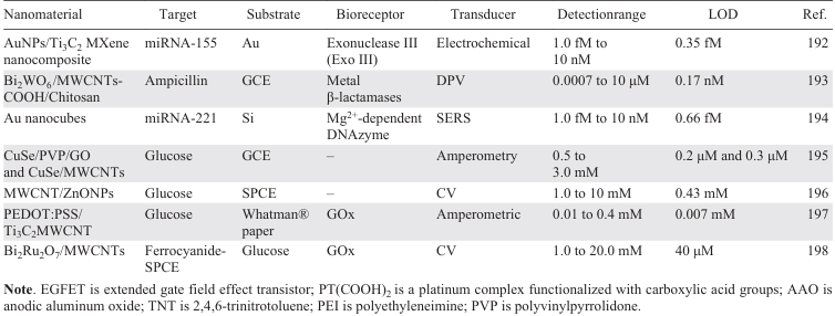
Overall, carbon-based materials, metal nanoparticles, transition metal dichalcogenides and quantum dots demonstrate tremendous potential in biosensor fabrication, offering high sensitivity, selectivity and versatility for diverse biomedical and environmental applications. Their enhanced properties contribute to superior sensitivity compared to other materials. Carbon-based materials, such as carbon nanotubes and graphene, offer a high surface area, excellent conductivity and biocompatibility, making them well-suited to electrochemical and optical biosensors. Metal nanoparticles, including AuNPs, PtNPs, and AgNPs, improve sensor performance by facilitating electron transfer and providing superior catalytic activity and signal amplification in optical and electrochemical detection platforms. TMDs have layered structures and tunable electronic properties, offering versatile applications in electrochemical and photoelectrochemical sensing. Integrating them with other nanomaterials improves signal transduction and lowers detection limits. QDs, with their size-dependent emission spectra, high fluorescence quantum yield and surface modifiability, enable highly sensitive and multiplexed detection in fluorescence and electrochemical biosensors. Together, these nanomaterials enhance the sensitivity, selectivity and reproducibility of biosensors, making them promising candidates for clinical applications. Table 2 summarizes recent advancements in nanomaterial-based biosensors, highlighting target analytes, bioreceptors, transduction techniques and detection performance.
The summarized data highlights the effectiveness of electrochemical and optical techniques using carbon-based nanomaterials, metal NPs, and 2D materials, which demonstrate superior sensitivity and selectivity in detecting diverse biomarkers. Electrochemical techniques are favored for clinical applications due to their simplicity and cost-effectiveness, as well as their ability to detect biomarkers with high sensitivity. Optical and field-FET-based methods also show great potential, particularly for POC diagnostics and real-time monitoring.
3. Applications in clinical diagnosis
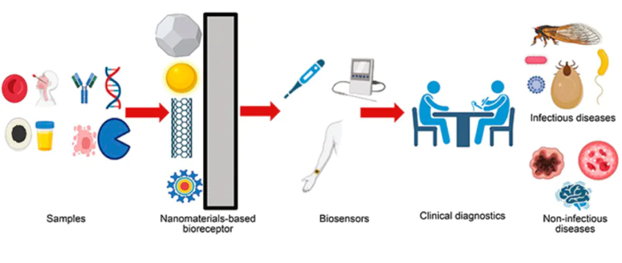
Nanomaterial-based biosensors have revolutionized clinical diagnostics by offering enhanced sensitivity and specificity for the rapid detection of a wide range of biomarkers and analytes. Such biosensors are increasingly being used for the early diagnosis of diseases such as cancer, cardiovascular disorders and infectious diseases, where the ability to detect low concentrations of biomarkers is crucial. For example, the use of AuNPs and CNTs in biosensors enables the ultra-sensitive detection of cancer-specific antigens and cardiac biomarkers, resulting in earlier and more accurate diagnoses. Nanomaterial-based biosensors are also used in POC testing, enabling real-time monitoring of glucose levels in diabetic patients and rapid pathogen identification in infectious diseases. The versatility of nanomaterials such as graphene and quantum dots in these biosensors also enables multiplexed detection, whereby multiple biomarkers can be identified simultaneously, thereby enhancing diagnostic precision further. In this section, we will explore the application of nanomaterial-based biosensors in clinical settings.
3.1. Non-infectious diseases biomarker detection
Biomarkers are measurable indicators that reflect the interactions between biological systems and potential hazards, aiding in disease diagnosis, prognosis, and treatment monitoring. They include nucleic acids, proteins, metabolites, and other biomolecules relevant to various diseases. Non-infectious disease biomarkers, particularly those associated with neurodegenerative diseases and cancer, are increasingly being detected using biosensors due to their high selectivity, sensitivity, and rapid detection capabilities.[199][200]
The development of electrochemical biosensors utilizing graphene derivatives, CNTs and AuNPs has greatly benefited cancer diagnosis. For example, an aptasensor was created to detect microRNA-128, a biomarker for acute lymphoblastic leukaemia.[201] This biosensor incorporated AuNPs, Fe3O4 and rGO to provide a large surface area for aptamer immobilization and enhance conductivity. The aptasensor demonstrated excellent specificity and sensitivity, with a LOD of 0.0055 fM in MB and 0.0053 fM in hexacyanoferrate respectively, rendering it a promising tool for early diagnosis and monitoring. Similarly, another aptasensor using an Au/graphene nanocomposite sensing platform and a CuS/graphene nanocomposite label was developed for the detection of leukaemia cancer cells.[202] This sensor could measure cell concentrations within the range of 50 to 10.000 cells mL–1 with an LOD of 18 cells mL–1 in blood samples. The catalytic activity of CuS/graphene increased the current of the biosensor in parallel with the addition of leukaemia cells, offering sensitive and reliable cancer detection. Laser-scribed graphene (LSG)-based electrodes have also been used to improve POC biosensing.[203] An LSG electrode modified with AuNSs successfully detected HER2 with a LOD of 0.008 ng mL–1, and this was integrated with a mobile application for real-time POC use. A mixture of thiol-modified DNA aptamer and mercaptohexanol was coated onto the LSG electrode surface. A BSA solution was then applied to reduce non-specific adsorption before the HER-2 sample was incubated on the aptasensor.
Early diagnosis of Alzheimer’s disease is crucial for treatment and prognosis. However, the complex composition, low content and individual differences of target proteins in real clinical samples still present challenges for the ultrasensitive and ultraspecific detection of early biomarkers for this disease. An ultrasensitive aptasensor using an RNA aptamer/AuNPs has been developed to detect amyloid beta (Aβ). The sensor can measure ultra-low concentrations of 0.4 pg mL–1.[204] Another aptamer was designed to detect tau381 in human serum samples. This was achieved by immobilizing thionin/carboxyl graphene/AuNPs on the GCE surface. This sensor exhibited a linear correlation with tau381 concentration ranging from 1.0 pM to 100 pM, with a LOD of 0.70 pM, thereby facilitating early-stage diagnosis.[205] Another aptasensor for Aβ40 was developed using TiO2/Au – C3N4 .[206] The sensor exhibited a detection range of 10−15 to 10−11 g mL–1 and an exceptionally low LOD of 0.33 fg mL–1. Similarly, a portable chemoresistive biosensor utilizing PPy NPs could detect both Aβ42 and Aβ40 with respective detection limits of 9.09 and 5.71 fg mL–1.[207] The development of the proposed Aβ sensing platform involves the following steps (Fig. 9a): (1) fabrication of a low-cost biochip using multiple interdigitated microelectrodes (IDμEs) on a single PCB substrate, (2) synthesis of PPy, its integration onto the IDμEs, and subsequent covalent immobilization of the bio-receptors, followed by analyte detection. The design and development of a portable read-out circuit, for current-voltage measurements (Fig. 9b) and the Android-based mobile application used for data acquisition and analysis (Fig. 9c) offer a cost-effective alternative to expensive brain imaging and provides a promising non-invasive method of diagnosis.
![[{"id":"adbpqvr6j_","type":"paragraph","data":{"text":"Schematic depiction of the Aβ sensing platform: (<i>a</i>) bioelectrode fabrication steps; (<i>b</i>) probe station setup for I – V (current-voltage) characterization of the sensor array; (<i>c</i>) pictorial representation of the portable readout platform. Reproduced with permission from the Ref. 207."}}]](/storage/images/resized/VWlYA0hGjaPqF6fthmxnuFdBNgMWhybx9cYJ99ei_xl.webp)
POC biosensors for glucose and other metabolic markers have also gained attention. Azimi et al.[208] developed miniaturized biosensors using vertically aligned CNT (VACNT) arrays for glucose detection in human plasma samples, achieving a LOD of 23 mM. Fig. 10a illustrates the fabrication steps where the silicon electrodes were treated with 10% HF to expose the VACNT contact layer, followed by electrochemically (EC) pretreatment with NaOH to add carboxyl groups for enzyme attachment. The arrays were then activated using EDC/NHS chemistry to covalently bind GOx. After applying the enzyme solution, the electrodes were washed with BSA-containing buffer. Another portable glucose biosensor integrating poly(norepinephrine)/Fe3O4/GOx/SPE with a smartphone analyzer, achieved a low LOD of 6.1 mM.[209] Fig. 10b illustrates the fabrication steps of the poly(norepinephrine)/Fe3O4/Gox modified SPE sensor for POC glucose detection. First, Fe3O4 NPs were synthesized by co-precipitation and subsequently coating with poly(norepinephrine) in tris(hydroxymethyl)aminomethane (TRIS) buffer. Second, GOx enzyme is immobilized on the Fe3O4@poly(norepinephrine) nanomaterial. Finally, Fe3O4@poly(norepinephrine)/GOx composite is deposited onto the SPE surface to fabricate the biosensor. Jędrzak et al.[210] further improved the sensitivity of glucose detection using a mobile-assisted POC biosensor based on βCD/Fe3O4/poly(norepinephrine)/GOx/SPE, achieving a LOD of 3.2 mM.Recently, an Ag-conjugated NiO NPs-on-wax screen-printed microfluidic paper-based colorimetric biosensor was developed for uric acid detection.[211] In this system, NiO/PVP/Ag NPs catalyzed the hydrolysis of uric acid, generating hydroxyl radicals, which subsequently oxidized 3,3',5,5'-tetramethylbenzidine, resulting in a distinct color change. The LOD of uric acid in simulated body fluids, phosphate buffer saline and deionized water was determined to be 0.03, 0.11 and 0.06 μM, respectively. rGO nanosheets and a cobalt nitride sensing platform immobilized with lactate oxidase were successfully used to detect lactic acid.[210] The introduction of rGO nanosheets, which have high electron-transfer kinetics and a large surface area, enhanced the current signal over the detection range of 1.0 to 80 mM, enabling an LOD of 0.51 mM to be achieved.[212] CdSe/CdS/ZnS nanocrystals enabled the optomagnetic detection of hErbB2 biomarkers in human serum,[213] providing a rapid, visually accessible diagnostic method.
![[{"id":"ROwxxqSxTK","type":"paragraph","data":{"text":"(<i>a</i>) Fabrication steps for the POC glucose detection system using VACNT arrays. Electron beam (EB) evaporation and radio frequency (RF) sputtering were employed to deposit catalyst and adhesion layers necessary for VACNT growth on the sensor substrate. Reproduced with permission from the Ref. 208. (<i>b</i>) Schematic illustration of the fabrication steps of the poly(norepinephrine)/Fe<sub>3</sub>O<sub>4</sub>/Gox modified SPE sensor for POC glucose detection. Reproduced with permission from the Ref. 209."}}]](/storage/images/resized/XPrU6Eh09vk7PihIAI9gCfDf1YeA2JlZ9pykUy91_xl.webp)
3.2. Infectious biomarker detection
The development of nanomaterials-based biosensors has significantly improved the specific and selective detection of the infectious agents responsible for various diseases, including hospital-acquired infections, foodborne illnesses, pandemics and water contamination. Several innovative biosensors have been developed for this purpose. One such example is a novel optical SPR-based bioanalyzer designed to detect infectious bursal disease virus (IBDV) with a detection limit approximately 1/18th that of traditional diagnostic strips.[214] For influenza detection, a boron-doped diamond electrode modified with polyclonal anti-M1 antibodies was employed, achieving a limit of 1.0 fg mL–1 of M1 protein in five minutes.[215] A polydiacetylene-based sensor functionalized with anti-H5 influenza antibody demonstrated sensitive detection of the H5 influenza virus with a LOD of 0.53 copies mL–1, providing visual confirmation via a noticeable color change.[216]
A microfluidic chip integrated with rGO was developed for label-free detection of H1N1 virus (Fig. 11).[217] The chip was fabricated using EDC/NHS chemistry to immobilize antibodies, achieving an improved LOD of 0.5 PFU mL–1, demonstrating high sensitivity for early virus detection. Chronoamperometric analysis showed that the current increased linearly with H1N1 concentration within the range of 1.0 to 104 PFU mL–1. Shen et al.[218] developed a biosensor based on CRISPR-Cas12a-powered magnetic NPs and QDs for detecting Salmonella, with a LOD of 86 CFU mL–1. This biosensor addresses limitations found in conventional CRISPR-based fluorescence detection methods, which often suffer from lower fluorescence quantum yields and higher background noise, thus limiting sensitivity and dynamic range. QDs were also combined with 2D Ti3C2 to produce a biosensor for SARS-CoV-2 and FluA detection, offering high stability, sensitivity and quick results within 20 min.[219] This biosensor has been shown to be more sensitive than the conventional AuNP-based ICA system when used with throat swab samples.
![[{"id":"JG05QJxKx7","type":"paragraph","data":{"text":"Schematic representation of a microfluidics-integrated electrochemical immune-sensing chip coated with rGO and subsequently undergoing antibody immobilization using EDC/NHS coupling for influenza virus H1N1 detection. Reproduced with permission from the Ref. 217."}}]](/storage/images/resized/WLSg475yDh5dwRqzR07WlE8wpLvgkfoWc9tBV1Pv_xl.webp)
Laser-scribed graphene (LSG)-based electrodes with 3D Au NPs were used to detect the SARS-CoV-2 spike protein, achieving a LOD of 2.9 ng mL–1 in clinical serum samples.[220] The sensor was designed by modifying the Au-LSG electrode using cysteine/EDC/NHS, followed by the immobilization of antibodies and antigens. The system was integrated into a handheld POC detection system operated using a custom smartphone application, providing a user-friendly diagnostic setup (Fig. 12). The results were compared with those from commercial RT-PCR, antibody blood tests and enzyme-linked immunosorbent assay (ELISA) IgG and IgA tests. The system offered an LOD of 2.9 ng mL–1, which is lower than that of conventional diagnostic systems. rGO-based FET biosensors were employed for the rapid and selective on-the-spot detection of the SARS-CoV-2 spike protein. These biosensors exhibited distinctive electrical behavior, characterized by measurable changes in the electrical conductance of the rGO channel upon binding of the spike protein. This label-free detection enabled a highly sensitive response with the LOD as low as approximately 1 fg mL–1. Additionally, the biosensor provided results in less than 2 minutes, considerably faster than conventional RT-PCR tests, which typically take several hours. The sensor also demonstrated excellent selectivity, showing minimal interference from other viral proteins or biological substances. These features highlight the potential of rGO-based FET biosensors as effective tools for rapid, accurate, and portable COVID-19 diagnosis at the POC.
![[{"id":"mPTUD8CSZ6","type":"paragraph","data":{"text":" (<i>a</i>) Photo of the portable, handmade potentiostat connected to a smartphone via a USB-C connection, recording the signal using customized KAUST software. (<i>b</i>) Close-up photo of the potentiostat on the device (scale bar: 1 cm). (<i>c</i>) Pinout diagram of the KAUST device. Reproduced with permission from the Ref. 220."}}]](/storage/images/resized/6UM8LUC2egZMdMiU1KuWojPzRqoN2JGDoT5izfR1_xl.webp)
A DNA aptasensor was developed [221] that can selectively bind to influenza A/H1N1 and SARS-CoV-2. This uses an Au nanopopcorn substrate to produce a signal via the surface-enhanced Raman scattering (SERS) method (Fig. 13). Raman Cyanine 3 and Rhodamine Red-X reporters were attached to the DNA aptamer terminals, producing strong SERS signals in the Au nanopopcorn substrate’s nanogap. The DNA aptamer is selectively detached from the substrate when the influenza A virus or SARS-CoV-2 approaches the Au nanopopcorn substrate, due to the significant binding affinity between the virus and the equivalent DNA aptamer. The SERS signal intensity decreases with increasing target virus concentration. The SERS-based sensing platform is a new diagnostic tool that can quickly discriminate between these two respiratory diseases, preventing their spread. The influenza A virus has been successfully detected at concentrations as low as 190 viral particles per mL using an SERS-based Ag nanoisland substrate.[222] These Ag nanoislands are nanostructured films composed of closely spaced silver nanoparticles deposited on a surface, resembling tiny «islands» under a microscope. Their morphology allows strong enhancement of the electromagnetic field at the surface, which amplifies the Raman signal of molecules near or attached to them. In this study, the Ag nanoisland SERS substrate was functionalized with specific antibodies against the influenza A virus, enabling selective binding of the virus particles. Upon binding, the interaction led to a significant enhancement in the Raman signal, allowing for highly sensitive and specific virus detection.
![[{"id":"3LI5SvvAkE","type":"paragraph","data":{"text":"Schematic illustration for the detection of influenza A/H1N1 and SARS-CoV-2 using the dual-mode SERS aptasensor. Two Raman reporters-labeled DNA aptamers are hybridized with capture DNAs on the Au nanopopcorn substrate. The internal standard 4-mercaptobenzoic acids (4-MBAs) are immobilized along with aptamer DNAs on the Au nanopopcorn substrate. Recognition of target protein induces the aptamer’s conformational change, leading to decreased corresponding Raman signal intensities. Reproduced with permission from the Ref. 221."}}]](/storage/images/resized/HVZQcU7uWHBbTOaLIaG42JLztHXlki6KbXTiKox9_xl.webp)
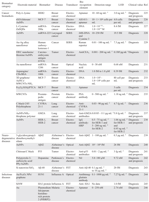
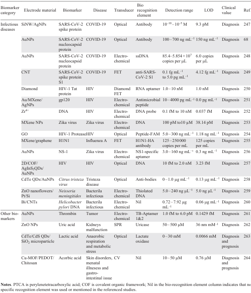
Paper-based and colorimetric biosensors have also been developed for the rapid detection of Zika virus and microRNA biomarkers, demonstrating remarkable stability and potential for widespread POC applications.[223][224] Table 4 summarizes the nanomaterial-based biosensors categorized by biomarker type, highlighting the most suitable techniques for specific biomarkers to help select optimal methods for clinical and POC applications. Electrochemical techniques remain the most widely used due to their high sensitivity, selectivity and reproducibility. Meanwhile, optical and FET-based biosensors offer promising solutions for rapid detection, making them ideal for real-time diagnostics.[225-245][246-264]
Although this review focuses on the clinical potential of nanomaterial-based biosensors, the examples discussed cover a variety of approaches with different levels of instrumental complexity, cost and feasibility for widespread diagnostic use. Some biosensors rely on sophisticated techniques such as SERS and SPR,[265][266] which require expensive, specialized equipment and therefore have limited applicability in settings with limited resources. In contrast, simpler electrochemical methods such as CV, EIS and DPV offer more accessible solutions for POC testing.[267] Differences in portability, cost and required expertise play a crucial role in determining the practical adoption of these technologies. Future research should focus on developing user-friendly, cost-effective biosensors that maintain high specificity and sensitivity, and are feasible for large-scale clinical deployment. By distinguishing between high-end, laboratory-based techniques and POC diagnostics, this review aims to provide a clearer perspective on the real-world applicability of emerging biosensor technologies.
In addition to developing selective and sensitive biosensors, practical factors such as assay time and long-term stability are essential. for their real-world application. Assay time is particularly important in clinical diagnostics, where prompt results are required. Although many biosensors enable rapid detection, reducing assay times without compromising sensitivity remains challenging. Nanomaterial-based platforms and signal amplification techniques can help to achieve this, thereby improving POC diagnostics.[268] The long-term stability of biosensor components is also essential for their commercial use. Stability can be impacted by factors such as humidity, temperature and degradation over time, but preservation approaches such as freeze-drying and encapsulation can enhance storage longevity, particularly in low-resource settings.[269]
4. AI-driven advancements in biosensors
Recent advancements in artificial intelligence (AI) have significantly enhanced the capabilities of biosensors, particularly with regard to real-time analysis, selectivity and sensitivity. For instance, machine learning algorithms have been incorporated into electrochemical biosensors to analyses complex signal patterns and achieve more precise measurement of analytes.[270][271]One study demonstrated the use of machine learning-assisted non-enzymatic electrochemical biosensors for urea detection, showcasing enhanced performance via the functionalization of CNTs with Cu2O micro-flowers.[272] AI has also been used to optimize the industrial production of CNT-based biosensors,[273] which can rapidly and label-free detect several important biomarkers. Furthermore, AI has facilitated the development of hybrid nanomaterials for use in biosensors. Integrating inorganic and organic nanostructures, including polymer-based nanomaterials, and metal NPs, has led to multifunctional systems that enhance biosensor performance. These hybrid materials enhance the sensitivity and selectivity of biosensors, particularly when it comes to detecting environmental contaminants and clinical biomarkers. AI-driven models have been optimized for electrochemical aptasensors. These models adjust for variations in environmental conditions, changes in the sample matrix, and sensor drift, resulting in improved reproducibility and stability of sensor responses.
Moreover, machine learning algorithms have been applied to non-invasive systems for continuous health monitoring, offering real-time data interpretation and improved disease treatment in the realm of wearable and portable biosensors.[274] AI has also played a transformative role in POC biosensing, where its integration with biosensor systems enhances accuracy and operational efficiency, particularly in low-resource clinical settings.[275] Together, these examples demonstrate the transformative impact of AI on biosensor technology, paving the way for more efficient, accurate and versatile diagnostic tools.
5. Challenges and future perspectives
In conclusion, advanced biosensors based on nanostructured materials represent a revolutionary approach to clinical diagnostics, offering significant improvements over traditional methods. While conventional diagnostic systems, such as ELISA or PCR, often require hours for analysis and have detection limits in the micromolar to nanomolar range, nanomaterial-based biosensors demonstrate ultrafast response times often under 10 minutes and sensitivities reaching down to the femtomolar or even attomolar levels. These exceptional features, enabled by materials such as graphene, CNTs, metal NPs, and QDs, facilitate the development of highly miniaturized, portable, and cost-effective devices. Such biosensors are especially advantageous for POC diagnostics and in low-resource environments, where rapid and accurate results are critical for effective clinical decision-making.
Although nanomaterial-based biosensors have great potential for clinical applications, they face several challenges that need to be addressed for them to be successfully translated into real-world diagnostic systems. One major challenge is achieving consistency and reproducibility in nanomaterial synthesis, as variations in morphology, surface chemistry and functionalization efficiency can result in inconsistent sensor performance. Automated synthesis platforms and standardized functionalization protocols can be employed to address this and ensure batch-to-batch consistency. Another limitation is the lack of commercially available, standardized nanomaterials, which hinders large-scale production. Designing reproducible, scalable and cost-effective synthesis protocols is therefore crucial. Another critical challenge is mitigating the effects of matrix interference and non-specific binding, which often impact the performance of biosensors in complex biological matrices such as blood, serum and urine. Advanced surface modification techniques, including PEGylation, self-assembled monolayers, EDC/NHS and nanomaterial coatings, can minimize these effects and enhance specificity. Furthermore, scaling up production while maintaining high functionality and quality remains a significant challenge, given that nanomaterials often require precise synthesis methods that are difficult to adapt for mass production. Addressing these challenges will require the development of more effective, scalable production methods and enhanced quality control measures.
Achieving high selectivity and sensitivity remains difficult, particularly in clinical samples. To address this issue, innovative molecular recognition elements with high binding affinities, such as antibody fragments, engineered aptamers and MIPs, are being explored. Additionally, hybrid nanomaterials, such as graphene--metal NPs hybrids, can enhance electron transfer and improve signal transduction, thereby improving the sensor’s overall performance. Signal amplification strategies also play a vital role in improving detection limits, particularly in samples with low concentrations. Techniques such as redox cycling, hybrid nanocomposite labels and enzyme-assisted amplification have shown promise in significantly enhancing biosensor sensitivity.
Moreover, integrating biosensors with AI and POC platforms can improve their reliability and efficiency in real-time clinical applications. Machine learning algorithms and AI-driven data analysis minimize errors, improve pattern recognition and facilitate the interpretation of complex datasets. Similarly, smartphone- and microfluidics-based platforms offer promising opportunities for decentralized and rapid diagnostics, particularly in settings with limited resources. Looking to the future, emerging technologies such as 2D nanomaterials, hybrid transducers and plasmonic nanostructures have significant potential to improve sensor performance. Furthermore, expanding the use of wearable biosensors for continuous monitoring paves the way for real-time, personalized medical care. These developments, coupled with their seamless integration into AI-based systems and POC platforms, are expected to transform clinical diagnostics and improve healthcare outcomes. However, overcoming challenges such as the toxicity of certain nanomaterials and ensuring biocompatibility is essential for their safe integration into clinical settings. Evolving production procedures, improving functionalization tactics and continuing to innovate in terms of AI integration and hybrid materials will enable these biosensors to overcome existing hurdles and pave the way for more competent, scalable and clinically applicable diagnostic tools.
6. List of abbreviations
AAO — anode aluminum oxide,
BSA — bovine serum albumin,
CDs — carbon quantum dots,
COx — cholesterol oxidase,
CV — cyclic voltammetry,
DPV — differential pulse voltammetry,
EDC/NHS — N-ethyl-N’-(3-dimethyl aminopropyl) carbodiimide/N-hydroxy succinimide,
EIS — electrochemical impedance spectroscopy,
ECL — electrochemiluminescence,
EFT — field-effect transistor,
ELISA — enzyme-linked immunosorbent assay,
GCE — glassy carbon electrode,
GEFT — graphene-based field-effect transient,
GQDs — graphene quantum dots,
GO — graphene oxide,
GR — graphene,
GOx — glucose oxidase,
ITO — indium tin oxide,
LSG — laser-scribed graphene,
LIG — laser-induced graphene,
LDH — lactate dehydrogenase,
LSPR — localized surface plasmon resonance,
LOD — low limit of detection,
MOF — metal-organic framework,
MIP — molecular imprinted polymer,
MWCNTs — multi-walled carbon nanotubes,
NPs — nanoparticles,
NWs — nano wires,
PCR — polymerase chain reaction,
PEDOT:PSS — poly(3,4-ethylene dioxythiophene):polystyrene sulfonate,
PEG — polyethylene glycol,
POC — point-of-care,
PPy — polypyrrole,
PTCA — 3,4,9,10-perylene tetracarboxylic acid,
rGO — reduced graphene oxide,
SPCE — screen-printed carbon electrode,
SPE — screen printed electrode,
SWCNTs — single-walled carbon nanotubes,
ssDNA — signal strand DNA,
SPR — surface plasmon resonance,
SERS — surface-enhanced Raman scattering.
References