Keywords
Abstract
Combination of the beneficial properties of gold nanoparticles and organic fluorophores forms the basis for the development of many advanced agents for bioimaging and laser medicine. The localized surface plasmon resonance of gold nanoparticles enables the control of the optical response and photodynamic activity of neighbouring molecules, being a powerful tool for fine tuning of their target physicochemical properties. Among numerous reviews devoted to the synthesis and optical properties of gold nanoparticles and their applications in particular areas of medicine and bioanalysis, there are no recent studies focusing on specific features inherent in hybrid structures of gold nanoparticles with fluorophores. Therefore, the goal of the present review is to consider the features of such plasmonic structures, the basic principles responsible for their optical properties, and modern approaches to attain multiplexed and multimodal response. The review also describes the existing combinations of gold nanoparticles and organic fluorophores and hybrid structures based on these components and presents their role for solving relevant tasks of biomedicine.
The bibliography includes 240 references.
1. Introduction
Theranostics, an advanced approach in medicine based on combination of diagnostics and therapy of diseases, poses new challenges to modern chemistry and materials science. In particular, it is necessary to prepare medicinal agents possessing an integrated action. A large area in the development of theranostic agents is occupied by noble metal nanoparticles, among which gold nanoparticles (GNPs) are especially relevant as most safe for biomedical applications The prospects of GNPs are determined by their unique physicochemical properties associated with the localized surface plasmon resonance (LSPR), which arises upon the interaction of metal nanoparticles with electromagnetic radiation in the optical range.
To solve the issues of theranostics, GNPs are most often converted to more complex hybrid structures, which makes it possible to expand their functional properties. Gold nanoparticles can be combined with various organic molecules,[1][2] polymers,[3][4] proteins,[5][6] or metal-organic frameworks;[7][8] a special place among these compositions belongs to the GNP combinations with organic fluorophores. In addition to the useful properties of GNPs, these hybrid structures provide the possibility of fine tuning of photophysical properties of the proper fluorophore, in particular, fluorescence quenching or enhancement and control of the photodynamic activity are possible.
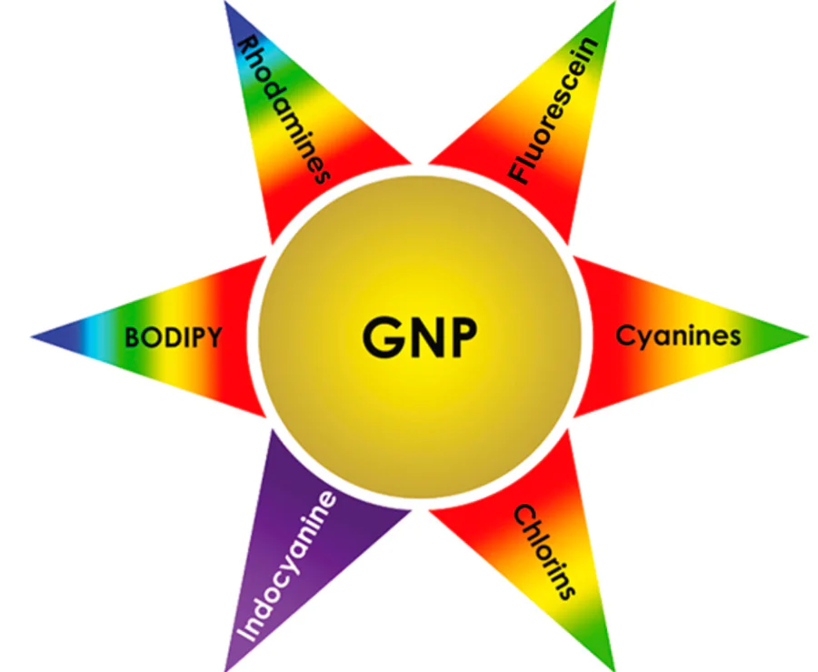
The applications of hybrid structures based on GNPs and molecular fluorophores are determined by their combined properties (Fig. 1). In view of the ability of GNPs to enhance the optical response of the fluorophore, design of the corresponding biosensors [9] and bioimaging probes [10][11] is underway. In particular, the concept of bioimaging probes is based on the idea that after predominant accumulation in or targeted transport to the affected tissues (typically malignant tumours), the probes produce the contrast in the sites of interest under laser irradiation during a diagnostic procedure or direct surgery. Owing to the pronounced photothermal effect of GNPs, the hybrid structures are promising for hyperthermia therapy. In this case, the presence of a fluorophore is important for monitoring the temperature of heating of the particles by means of fluorescence ratiometry.[12-14] The local temperature monitoring is necessary to prevent overheating of tissues, which would harm the nearby healthy cells. Finally, GNPs and structures based on them can be functionalized rather easily by vector molecules and, hence, they can act as carriers for targeted transport of drugs [15][16] or genes.[17][18] A fluorophore incorporated in a nanoplatform of this type serves as a tool for optical control of the targeted delivery facilitating local release of the active agent.
![[{"id":"RurcUN9gjq","type":"paragraph","data":{"text":"Biomedical applications of hybrid structures based on gold nanoparticles and fluorophores determined by their physico-chemical properties."}}]](/storage/images/resized/bfhGEqAb2ZARxpuzy3lx7dl1LgNw7eEjMY79OUha_xl.webp)
Owing to their biomedical potential, hybrid structures based on GNPs and organic fluorophores attract a lot of research interest, which is manifested as numerous experimental and theoretical studies published on this subject. However, among the publications, the number of review papers providing systematization of the synthesis methods of photoactive plasmonic molecular structures and giving critical analysis of their properties, benefits, and application challenges is moderate. The vast majority of reviews focus on either GNPs, including methods of their synthesis [9-21] and tuning of their optical properties,[22][23] or to their use in particular fields of bioanalysis and medicine.[24-28] The goal of this review is to address hybrid structures based on GNPs and organic fluorophores, including description of the fundamental principles of their optical response, preparation methods, and the most outstanding examples of biomedical testing.
2. Plasmonic properties of gold nanoparticles: origin and associated physicochemical effects
2.1. Localized surface plasmon resonance phenomenon
The localized surface plasmon resonance is a very complex phenomenon related to surface physical chemistry and chemical physics. LSPR is commonly considered as oscillations of charge density on the surface of metal nanoparticles that resonate with an external electromagnetic radiation. A schematic image of the LSPR effect is depicted in Fig. 2. The first theoretical description of LSPR was proposed in the early 20th century by Mie.[29] Since then, analytical expressions for the field excited by a planar monochromatic electromagnetic wave have been derived for nanostructures of various sizes and shapes and for various types of materials; they can be found, for example, in monographs.[30][31]
![[{"id":"I6jjRLK4q4","type":"paragraph","data":{"text":"Schematic illustration of LSPR on the surface of gold nanoparticles"}}]](/storage/images/resized/pPAM7hoa4Ldu8po8bzQJH11xixF2jblktAs56EVD_xl.webp)
Localized surface plasmon resonance is a local effect, and the electromagnetic field generated due to LSPR is rapidly decreased. In the simplest case of a metal sphere of diameter D, the field of a dipole excited by an incident electromagnetic wave decreases with increasing distance R from the fluorophore to the centre of the sphere proportionally ~(D/[0.5 D + R])3.[32] The enhancement factor of the local electromagnetic field ~|E|2/|E0|2 is responsible for increasing intensity of the fluorescence of molecules; therefore, it varies inversely proportional to the sixth power of R. Modern numerical simulation methods provide a fairly accurate description of the electromagnetic field distribution near plasmonic nanoparticles, consistent with the experimentally observed dependences. For example, calculations performed by Zar’kov et al.[33] for spherical gold nanoparticles with a diameter of 60 nm showed that the range of variation of |E|2/|E0|2 in the localization region confined by a 5-nm thick radial layer is 1.5 – 37.8 in the plane with the most inhomogeneous electromagnetic field. The cited publication also gives details of estimation of electromagnetic field enhancement factors for gold nanorods with various aspect ratios.
The localized surface plasmon resonance accounts for certain physicochemical properties of noble metal nanoparticles, which underlie some of their applications. In particular, a characteristic intense band corresponding to the resonance absorption of electromagnetic radiation by a nanoparticle appears in the absorption spectra. One more consequence of LSPR is the enhancement of various modes of optical signals from molecules located directly on or near the surface. The best-known example is surface-enhanced Raman scattering (SERS). The SERS effect consists in the increase in the Raman cross-section of a molecule, which can be as high as 12 orders of magnitude.[34] The so-called hot electrons excited in a metal nanoparticle due to LSPR can act as catalysts in some chemical photoreactions that take place on the metal surface.[35]
2.2. Effect of plasmon resonance on the fluorescence of adsorbed molecules
The local electromagnetic field on the surface of a plasmonic nanoparticle naturally affects all optical processes occurring at or near the interface, including fluorescence of molecular fluorophores. Currently, the theory of fluorophores located near plasmonic nanoparticles has been described at a considerably in-depth level and can be found in monographs.[29][36] In this review, we address only basic issues needed to understand the general principles of the optical response of hybrid molecular plasmonic nanostructures.
Since fluorescence includes two stages, excitation of a molecule and the subsequent emission of energy by this molecule, the local electric field of a nanoparticle can modify both processes and influence the energy transitions in the fluorophore both at the absorption wavelength λabs and at the emission wavelength λem. The rate constant for fluorophore excitation ke in an electric field of the nanoparticle at the absorption wavelength λabs is given by[32]
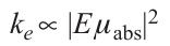
where μabs is the dipole moment of the transition corresponding to radiation absorption.
In comparison with the electric field of the incident electromagnetic wave E0, the local electric field E is enhanced due to LSPR; hence, the rate constant for fluorophore excitation also increases
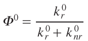
After excitation, the fluorophore molecule can undergo radiative or non-radiative decay of the excited state. For a free fluorophore, the former type of decay is characterized by the rate constant kr0, while the rate constant for the latter type of decay is knr0 . The fluorophore quantum yield in the absence of a plasmonic surface is expressed by the relation
The radiative stage of fluorescence of adsorbed molecules is modified in a more complex manner due to competition of two major effects: plasmonic enhancement and Förster resonance energy transfer (FRET). The key factor for the predominance of a particular effect is the distance between the molecule and the metal surface. In general, the influence of plasmonic nanoparticles on the excited state decay in a fluorophore can be described on the basis of Maxwell equations and FRET theory in which a molecule and a nanoparticle are considered as interacting dipoles.
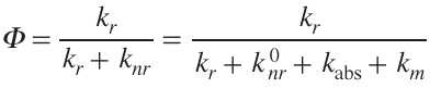
In a local electromagnetic field, both rates of the excited state decay in the fluorophore change to kr and knr, respectively. The rate constant for the radiative decay of the excited state knr increases due to the Purcell effect consisting in an increase in the emission rate of an oscillator in an inhomogeneous medium.[37] The rate constant for the non-radiative decay of the excited state knr = knr0 + kabs + km also increases, since new contributions to the rate constant appear, that is are, kabs, caused by the thermal dissipation of energy, and km, related to coupling to non-radiative electromagnetic modes.[38] Thus, the quantum yield of a fluorophore under LSPR conditions can be written as [38]
In view of the fact that the influence of LSPR on the rate constant of non-radiative decay of the fluorophore excited state knr has an intricate pattern, it is obviously very difficult to accurately describe the dependence on the distance to the surface. However, considering fluorescence quenching in terms of FRET theory makes it possible to follow important features inherent in plasmonic substrates. The accumulated experimental data indicates that the dependence of the energy transfer rate on the distance between the donor and the acceptor in the case of metal nanoparticles is described by Eqn (5), where n = 3 – 4.[39][40] Thus, this dependence differs from that typical of a molecular donor and acceptor pair in which the exponent is six.
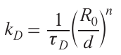
where tD is the fluorescence lifetime in the presence of an acceptor (nanoparticle), R0 is the distance at which the energy transfer efficiency is 50% (similarly to the Förster radius), d is the distance between the fluorophore and the nanoparticle.
Generally, higher energy transfer efficiency is inherent in a fluorophore – nanoparticle pair, which gave rise to the concept of ‘super-quenching’, known in the literature as the nanosurface energy transfer (NSET).[41][42] The NSET effect forms the basis for many analytical procedures operating by either activation or deactivation principle (decrease or increase in the fluorescence intensity of a signalling molecule, respectively) and responsible for a significant portion of practical applications of gold nanoparticles in the field of bioanalysis.[43][44]
In the case of small distances between a molecule and a surface, the metal-modulated rate constant for the non-radiative decay of the excited state knr reduces the excited state lifetime of the fluorophore tD = (kr + knr)–1 and decreases the quantum yield F. However, as the distance between the surface and the molecule increases, the rate of non-radiative decay of the excited state in the fluorophore decreases more rapidly than the radiative decay rate, which accounts for the increase in the quantum yield accompanied by switching of fluorescence quenching to fluorescence enhancement. When the distance exceeds the region of LSPR, the emission from the fluorophore no longer interacts with the local electromagnetic field.
Thus, relying on the views on fluorescence enhancement by a local field, the electromagnetic enhancement can be considered as the interaction of plasmon resonance with the excitation and emitted radiation. Hence, the following expression can be derived:[45]
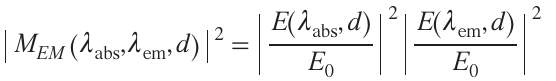
where |MEM|2 is the electromagnetic fluorescence enhancement factor.
An additional parameter that may contribute to the fluorescence enhancement is the directionality factor f of a fluorophore emission that interacts with LSPR. The parameter f depends on the polar and azimuthal angles of the excited state radiative decay; a detailed description of this dependence can be found in the literature;[46][47] here, we only recognize the fact that this dependence exists. Generally, the total fluorescence enhancement, characterized by the F/F 0 ratio, may be caused by any of three factors, or by their cumulative action, that is, the increase in the excitation rate constant ke, the increase in the quantum yield F due to the growth of kr, and the increase in the emission directionality f:[32]
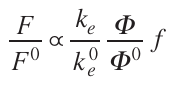
where F/F 0 is the fluorescence enhancement ratio relative to the fluorescence measured in the absence of metal nanostructures (e.g., free fluorophore molecules in a homogeneous aqueous medium).
It is noteworthy that the fluorescence enhancement factor F/F 0 appreciably depends on the orientation of the fluorophore molecule. Since the fluorophore molecules are usually randomly oriented, the enhancement factor measured for an ensemble of emitters is averaged over all of the possible orientations of the absorption and emission dipole moments mabs and mem.
Apart from the distance between the molecule and the surface, the spectral overlap of the LSPR absorption band of a nanoparticle and the excitation and emission bands of the fluorophore is also an important factor, determining the degree of coupling of all spectral processes and NSET efficiency. A simplified energy diagram and illustrative view of the major factors influencing the fluorophore emission near the plasmonic surface are depicted in Fig. 3.
![[{"id":"Pn3lxQYage","type":"paragraph","data":{"text":"(<i>a</i>) Energy diagram of excitation and decay of the excitation state of the fluorophore under LSPR effect and in a free space; (<i>b</i>) illustration of the fluorescence intensity of molecules located at different distances from the plasmonic surface; (<i>c</i>) spectral overlap of the absorption region of plasmonic nanoparticles with the absorption and emission regions of the fluorophore where all fluorophore kinetic parameters are influenced by LSPR [on the example of the absorption spectrum of gold nanorods with an aspect ratio of 2.1 (orange) and absorption (pink) and emission (lilac) spectra of cyanine 5.5]."}}]](/storage/images/resized/L0kIGDggWmyfpStF61JeOtoYePD5NLbKVaRHJgR0_xl.webp)
For a fluorophore subjected to LSPR of a nanoparticle, the fluorescence lifetime changes (most often, decreases). Evidently, this effect is also highly dependent on the distance between the molecule and plasmonic particle; the shorter the distance, the stronger the effect. There are a few elegant studies in which the lifetimes of fluorophores located at different distances from the metal surface were determined by using linkers of various lengths [48] or by using coatings of variable thickness.[49][50] Table 1 summarized published data on the fluorescence enhancement factors and fluorophore lifetimes in hybrid structures.
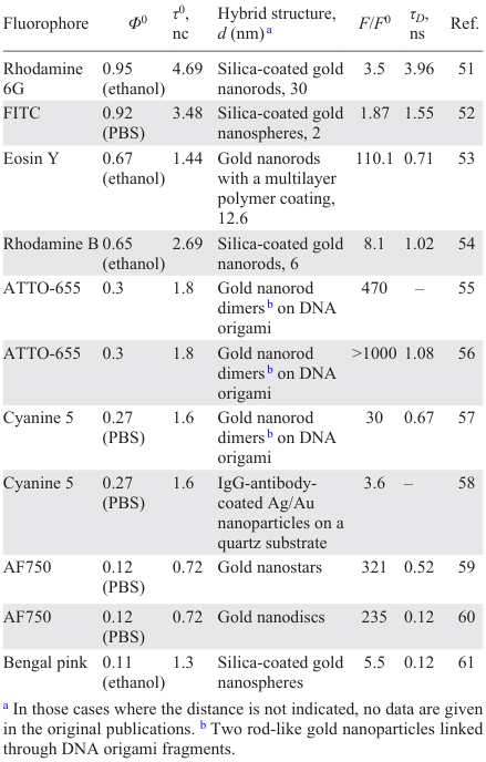
As can be seen from the Table, fluorescence enhancement varies from several times to several orders of magnitude. The highest enhancement factors are provided by GNP dimers. A more pronounced fluorescence enhancement appears owing to the known hot spot effect, which consists in the superposition of local electromagnetic fields in the vicinity of closely located plasmonic particles. In general, a more pronounced fluorescence enhancement is observed for molecules with moderate intrinsic fluorescence quantum yield.
2.3. Photothermal effect of gold nanoparticles
The ability of gold nanoparticles to scatter energy as heat upon absorption light, i.e., to exhibit a pronounced photothermal effect, is related to several factors. The major factor is LSPR, since the generated electromagnetic field, first of all, allows nanoparticles to convert light energy to thermal energy. One more important factor is the high heat conductivity of gold; in particular, in ensures heat transfer not only within the particle, but also to the environment. An additional factor is the high surface area to volume ratio generally characteristic of nanoparticles compared to micro and macro items. The combination of the above properties determines the potential of gold nanoparticles as agents for photothermal therapy (PTT).
A macroscopic model considering the generation and dissipation of heat by an aqueous suspension of gold nanoparticles in a measuring cell was proposed for calculating the efficiency of photothermal conversion.[62] The change in the temperature upon irradiation of a GNP dispersion is described by the energy balance equation [63]
where mi and Ci are the mass and the specific heat capacity of each i-th component of the physical system (nanoparticles, deionized water, cell), Eabs is the heat induced by absorption of electromagnetic radiation by the particles, Eloss is the heat lost to the environment.
If the total mass of the particles is insignificant compared to the mass of water and the cell and the specific heat capacity of the particles is low compared to the heat capacity of water and cell material, then the mass and heat capacity of the whole dispersion can be taken to be equal to the mass and heat capacity of water. Then the last two terms present in relation (8) can be expressed in the following way:[63]
where P is the power of the incident continuous laser radiation, Aλ is the absorbance of a nanoparticles dispersion that fills the cell volume at the wavelength λ, η is the photothermal conversion efficiency, h is the heat transfer coefficient, S is the surface area covered by the GNP dispersion, T0 is the ambient temperature.
Thus, the above expression can be converted to the following form:[63]
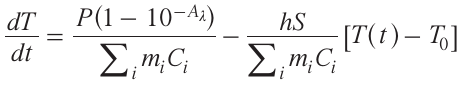
To simplify this equation, parameters A and B are introduced:
A is the energy absorption rate, B is the heat dissipation rate.
Then relation (13) acquires the form
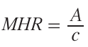
The conversion of electromagnetic radiation to heat by gold nanoparticles is characterized by two values: photothermal conversion efficiency η and the molar heating rate (MHR). The photothermal conversion efficiency depends mainly on the rate of absorption of light energy by the system (A) and is described by the expression [63]
The parameter MHR can be interpreted as the rate of the change in the temperature of the system normalized on the molar concentration of the heaters (in this case, nanoparticles).
The photothermal conversion efficiency η requires experimental determination by measuring the temperature variation with time during irradiation (А ≠ 0) and subsequent cooling of the nanoparticle dispersion (A = 0) and by finding the A and B values. Measurements of this type can be performed with a reasonable accuracy using terahertz spectroscopy, which has certain advantages over the methods using thermistors or methods based on infrared or luminescence thermometry. The photothermal conversion efficiency was determined by terahertz spectroscopy [63] for various types of gold nanoparticles and was found to reach 90% for 10 × 41 nm nanorods (Fig. 4).
![[{"id":"oceFvurdNi","type":"paragraph","data":{"text":"(<i>a</i>) Thermal image of an aqueous dispersion of gold nanorods demonstrating temperature distribution after laser treatment; (<i>b</i>) calculation of the light energy absorption rate A by a dispersion of gold nanorods by fitting the experimental dependence of the dispersion temperature on the irradiation time.<sup>63</sup>"}}]](/storage/images/resized/GPOKA6bzdcEsBpjuU9rlPzXwREdGJgZ5ePVx1M7s_xl.webp)
2.4. Dependence of optical and biological properties of gold nanoparticles on their size, shape, and surface chemistry
The particle size and morphology influence all properties of GNPs, including optical absorption and interaction with biological objects. In the simplest case of spherical nanoparticles, LSPR is manifested in the absorption spectra as a single band, the position of which is determined by particle diameter. In the case of small GNPs of 5 nm in diameter, the LSPR peak is in the range of 514 – 517 nm.[64][65] As the GNP size increases, absorption shifts to longer wavelengths, and for the largest particles with a size of 200 – 300 nm, the absorption maximum is in the 650 – 720 nm range.[66-68] Anisotropic GNPs have a more intricate absorption profile. Gold nanorods represent the most remarkable example. They exhibit two LSPR modes, which are commonly called transverse and longitudinal modes, caused by oscillations of conduction electrons along the long and short axes of the nanoparticle, respectively (Fig. 5).[69][70] Depending on the aspect ratio, the positions of both LSPR modes change, and the longitudinal mode may shift by hundreds of nanometres. The absorption spectral patterns of GNPs that have a few bulges, cavities, or other surface curvature elements (nanostars, nanocages, etc.) are even more complicated. The number of surface curvature elements with different geometric sizes determines the number of wavelength peaks on which LSPR is activated. For example, an imaginary nanostar with twenty completely separated arms of different lengths should give rise to twenty different LSPR absorption bands. Since structures of this type with numerous different single surface curvature elements cannot be obtained in practice and there are plasmon coupling effects, the absorption profile of nanostars, nanoflowers, nanourchins, and other similar particles is usually a common broad envelope curve resulting from the superposition of several LSPR bands (see Fig. 5). Thus, by controlling GNP shape, it is possible to shift the position of LSPR over a broad spectral range up to ~ 1900 nm,[71] i.e., to not only the first, but also the second transparency window of biological tissues, which further increases the biomedical potential of GNPs.
![[{"id":"Hj6q2I2JlV","type":"paragraph","data":{"text":"(<i>a</i>) Transverse and longitudinal LSPR modes in the absorption spectrum of 31.8 × 16.1 nm gold nanorods and results of simulation of electricfield distribution near the surface upon excitation with a field directed along the long or short axis.<sup>69</sup> (<i>b</i>) Comparison of the simulated absorption cross-section spectrum of the model of a perfectly symmetrical 50-spike nanostar (orange curve) and the experimental absorption spectrum of synthesized gold nanostars with a similar mean sizes (blue curve). The figures on the right of the coloured scale correspond to the relative strength of the local electric field.<sup>70</sup> Figure reproduced with permission from the American Chemical Society."}}]](/storage/images/resized/5NG7VMVXAJ0qWFgN3di3Rs3R84ilFu1XuFjSBKtz_xl.webp)
In the study of GNP biocompatibility, the central issue is the uptake of GNPs by model living objects (cells, bacteria, multicellular organisms) followed by evaluation of the metabolic activity. In early studies, attempts were made to find a correlation between the GNP toxicity and size. Some researchers concluded that a decrease in the particle size leads to increasing toxicity.[72][73] However, some other publications note, on the contrary, a higher toxicity of large GNPs compared to GNPs with smaller diameters.[74][75] The contradictions between the results are evidently attributable to multifactorial nature of the problem, which hampers drawing general conclusions concerning the correlation between GNP toxicity and size.
The subsequent studies were increasingly focused on evaluation of the toxicity for GNPs of various shapes, and they did not identify an unambiguous relationship either. Comparative studies of spherical and rod-shaped nanoparticles indicate that the latter are more toxic almost in all cases,[76][77] although in some studies no difference was identified.[78] However, comparison of spherical and so-called branched particles such as nanostars, nanoflowers, and nanocages brought the same result: branched particles always prove to be more toxic.[79][80] The higher toxicity of branched GNPs is probably due to two factors: their extensive surface area, which provides a larger contact area, and their ability to cause more damage to the cell because of their shape.
Although the size and shape of GNPs undoubtedly have some influence on the toxicity, the scientific community is now increasingly inclined to believe that the main factor determining the cellular uptake and toxicity of GNPs is their surface chemistry.[81][82] An illustrative study [83] addresses the biocompatibility of equal-size gold nanospheres with different surface charges and different compositions of adsorbed functional groups with the gram-positive Bacillus and gram-negative Shewanella bacteria. The authors used three types of particles with different coatings: anionic coating based on 3-mercaptopropanoic acid (MPA-AuNPs), cationic coating based on 3-mercaptopropylamine (MPANH2-AuNPs), and cationic coating based on a polyelectrolyte, polyallylamine hydrochloride (PAH-AuNPs). The particle toxicity was evaluated by the colony counting method and by respirometry, that is, measurement of the oxygen uptake by bacteria, which characterizes their viability (Fig. 6). The negatively charged MPA-AuNPs had the lowest toxicity. The incubation of both types of bacteria with MPA-AuNPs at concentration of 5 μg mL–1 on gold induced no toxic effect. In the case of particles with positively charged surface, both the concentration and particular bacterial species were significant. According to the colony counting method, MPANH2-AuNPs exhibited no toxicity against Shewanella at concentrations of up to 5 μg mL–1, but had a noticeable toxicity against Bacillus (less than 80% of cells remained viable) at a concentration of 0.05 μg mL–1. PAH-AuNPs at the same concentration proved to be toxic for Shewanella and very toxic against Bacillus (less than 10% of cells were viable). The higher toxicity of polyelectrolyte-coated particles was attributed to their higher surface charge density, which allows them to be better attached to bacteria. The high toxicity of both types of particles with the positively charged surface against Bacillus was attributed to the absence of the second lipid membrane in these bacteria.
![[{"id":"yx1CXTFXQl","type":"paragraph","data":{"text":"The left panel shows the overall experimental concept for evaluation of the toxicity of gold nanoparticles with different coatings against gram-positive and gram-negative bacteria. The right panel shows experimental data: (<i>a – d</i>) viability of the <i>Shewanella</i> (black) and <i>Bacillus</i> (grey) bacteria after incubation with MPANH<sub>2</sub>-AuNPs and PAH-AuNPs determined by the colony counting method. The white columns correspond to the viability of bacteria determined in the presence of an equivalent concentration of the polymer but without GNPs; (<i>e, f</i>) results of the respirometric analysis of <i>Shewanella</i> (<i>e</i>) and <i>Bacillus</i> ( <i>f</i>) incubated in the medium without (black) and with (red) MPA-AuNPs; the concentration was 5 μg Au mL<sup>–1</sup>. The oxygen uptake curves show a minor influence of MPA-AuNPs on the metabolic activity of the bacteria. The diagrams (<i>a – d</i>) show the p-values for the statistical significance in the independent t-test, ****p < 0.0001 (statistically significant values), ***p < 0.001 (values of high statistical significance), **p < 0.01 (values at the border of statistical significance).<sup>83</sup> Figure reproduced with permission from the Royal Society of Chemistry."}}]](/storage/images/resized/CWpUwW699rtWcrJnjVNXl8nfolO6iJxdtQfdMbVh_xl.webp)
A similar experiment was carried out by Vales et al.,[75] who divided gold spheres of 5 and 20 nm in diameter into three types according to the chemical nature of their surface groups: carboxylated (negatively charged), aminated (positively charged), and polyethylene glycol (PEG)-coated (neutral) surface. The surface-aminated particles had the highest toxicity against the BEAS-2B bronchial epithelial cell line irrespective of the size. The incubation with these particles at a concentration of 25 μg Au mL–1 resulted in a death of most cells. The carboxylated and PEG-stabilized particles proved to be non-cytotoxic up to gold concentration of 250 μg mL–1. In this case, the particle diameter did not significantly affect the result either.
In general, the elucidated, often intricate, effect of the GNP size, morphology, and surface chemistry on the properties indicates that the selection of particles for the design of biomedical agents should involve an integrated consideration of their parameters. However, it is obvious that anisotropic GNPs in which LSPR falls within the transparency window of biological tissues and those coated with biocompatible materials should be preferred for the photomedical purposes.
3. Hybrid structures based on gold nanoparticles and organic fluorophores
3.1. Organic fluorophores used in the hybrid nanostructures
Fluorophores suitable for the design of GNP-containing hybrid structures should possess photostability and exhibit high quantum yields. If hybrid nanostructures are intended for biomedical applications, it is highly important that the excitation and emission bands of the fluorophore be in the red or near-infrared region of the electromagnetic spectrum (650 – 900 nm). A serious issue is safety of their use. If nanostructures are bound to fluorophore in the last stage by immobilization on the souter surface, then after entering the body, the molecules will be in direct contact with the medium including blood and tissues. Fluorophores often contain functional groups able to undergo undesirable reactions. To avoid this, various micelles and polymer shells that protect the fluorophore and do not prevent emission are fabricated.[84][85] Apart from synthetic organic fluorophores,[86] other possible components of hybrid structures that endow them with optical response may include natural dyes isolated from plants or biotissues,[87-91] fluorescent proteins,[92] lanthanide-doped oxide nanoparticles,[93][94] and carbon quantum dots.[95] Of particular interest are metal nanoclusters with a countable number (usually not exceeding 30) of heavy atoms formed on amino acids, proteins, or DNA, which are by themselves small hybrid structures with high biomedical potential [96-98] and can also be incorporated into GNP-based plasmonic nanostructures.[99][100] The traditional synthetic dyes that are used most often in modern theranostic agents in combination with GNPs are summarized in Table 2.
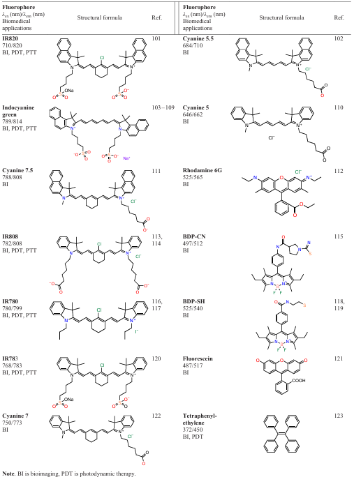
Indocyanine green (ICG) is obviously the most popular organic fluorophore for biomedical applications; importantly, it was recognized to be safe for use for therapeutic purposes. Indocyanine green is very convenient for use, since it is amphiphilic and negatively charged; it has a good response in the IR range and causes few allergic reactions. In addition, it has a short half-life in blood (150 – 180 s) and is excreted exclusively by the liver.[124] Molecules of indocyanine green rapidly and almost completely bind to blood proteins, thus being less prone to aggregation and increasing the quantum yield. The hydrodynamic radius of the resulting structures is comparable with the size of bound proteins, which is important for ICG transport and retention in lymph nodes.[103] In addition, ICG possesses a proven photodynamic action.[125]
A large family of fluorophores for theranostics is represented by other polymethine dyes: cyanines and squaraines. Their benefits include narrow and intense excitation and emission bands in the far optical range, as well as great variability of possible structure optimization. The molecules of cyanines have several sites for possible modification. The introduction of various groups into a polymethine dye molecule can provide a reasonable solubility in water, high photostability, and excitation and emission wavelengths appropriate for fluorescence imaging.[126-128] Like polymethines, dyes based on boron dipyrromethene (BODIPY) can be easily modified owing to the presence of three modification sites.[129] These fluorophores are distinguished by high quantum yields, photostability, and chemical stability.[130] High variability is inherent in the family of xanthene dyes, which includes important fluorescent agents such as rhodamines and fluorescein (FAM).
Special mention should be made of the fluorophores that exhibit aggregation-induced emission (AIE) effect consisting in the increase in the emission intensity upon the aggregation of molecules. Most often, AIE fluorophores are highly conjugated symmetrical compounds; tetraphenylethylene can be considered as an example.[123] The benefits of AIE fluorophores are due to the fact that they enable operation in any concentration range, being organic analogues of quantum dots. Fluorophores of this type are even regarded as new-generation agents for biosensors and imaging.[131]
The photophysical properties of many fluorophores considered above can be improved or expanded by incorporating them into hybrid structures based on gold nanoparticles. As mentioned above, GNPs can enhance the fluorescence signal of adsorbed molecules, increase the photodynamic activity (e.g., in the case of ICG), or can serve as scaffolds for dyes with aggregation-induced emission effects.
3.2. Preparation methods of hybrid structures based on gold nanoparticles and organic fluorophores
There are various approaches to the fabrication of hybrid structures based on gold nanoparticles and organic fluorophores. Conceptually, they can be divided into two groups according to the way of fluorophore immobilization on the GNP surface or in the nanoparticle shell: adsorption deposition and chemical conjugation (Fig. 7). The adsorption deposition of fluorophores from solutions is usually driven by one or several factors: (1) hydrophobic interactions; (2) electrostatic interactions; (3) formation of Au – S or Au – N bonds with the surface (characteristic of thiols and amines).
![[{"id":"eb-ZHSA6sq","type":"paragraph","data":{"text":"Strategies towards the fabrication of hybrid structures based on GNPs and fluorophores: (<i>a</i>) adsorption deposition controlled by hydrophobic (in relation to fluorescent-labelled protein), electrostatic, or specific interactions; (<i>b</i>) covalent conjugation through various functional groups; (<i>c</i>) key chemical reactions used for the conjugation of fluorophores to obtain hybrid structures. The colour of lines in part (<i>b</i>) corresponds to the colour of the reaction number in part (<i>c</i>)."}}]](/storage/images/resized/ool2Oijfpzm6FnDlWrqTYJGeANz1qspxr665ODbg_xl.webp)
The adsorption deposition is simpler than the chemical conjugation, but provides a lower efficiency of attachment of fluorophore molecules to the GNP surface. The covalent conjugation makes it possible to attach a larger number of molecules to the surface; it is more specific, but may lead to significant loss of fluorescence response due to generation of new channels for non-radiative energy transfer. Most often, the chemical conjugation of fluorophores to nanoparticles occurs via a linker and requires preliminary surface functionalization with small reactive molecules or polymers.[132] [133]
Among the methods of covalent conjugation of fluorophores to GNPs, the carboxylation-amidation reaction (reaction 2 in Fig. 7c) with N-hydroxysuccinimides is used most often. This method requires preliminary modification of the GNP surface with primary amino groups or carboxyl groups, depending on which functional group is present in the dye to be attached. Other commonly used reactions are azide – alkyne cycloaddition (reaction 3), reaction of thiols with maleimides (reaction 1), reaction of hydrazines with carbonyls (reaction 4), and the inverse electron demand Diels–Alder reaction (reaction 5). The methods for covalent conjugation of molecules to the GNP surface have been studied in detail recently, in particular, the dependences of the conjugation efficiency on the molecule nature, pH of the solution, temperature, and on a number of less obvious parameters such as stirring intensity have been elucidated.[134-136]
Special mention should be made of the method for the preparation of GNP-based hybrid structures called ‘DNA origami’. This approach gives ordered structures consisting of DNA chains and GNPs distributed in them, being attached to strictly definite nucleotides. If fluorescent-labelled DNA molecules are used to fabricate the hybrid structure, the process gives photoactive structures in which fluorescence can be enhanced 5000-fold.[137-141]An important feature of the structures obtained by the DNA origami method is the possibility of precision control of the fluorophore position relative to one or several gold nanoparticles.[142][143] Therefore, the fluorophore can be positioned in the region with the most pronounced enhancement of electromagnetic field, in particular, in so-called hot spots, that is, areas between the closely spaced plasmonic nanoparticles in which superposition of local electromagnetic fields takes place (Fig. 8).
![[{"id":"SrZ5O0GzuU","type":"paragraph","data":{"text":" (<i>a</i>) Sketch view of a hybrid structure in which two gold nanoparticles of 100 nm in diameter are attached to a columnar DNA origami containing the ATTO 647N fluorophore incorporated in the middle of a 6-helix bundle and thus positioned at the centre of the interparticle gap. (<i>b</i>) Numerical simulation of the electric field intensity in the equatorial plane of the dimer structure with an interparticle distance of 12 nm at a 640 nm wavelength and at the incident electric field polarization parallel to the dimer orientation.<sup>143</sup> Figure reproduced with permission from the American Chemical Society."}}]](/storage/images/resized/Yg2riuIBopuUbtPSsISSG848U86cG8ImazbU4A2I_xl.webp)
4. Fluorescent mono- and multimodal gold nanoparticle probes in bioimaging and cell biology
The hybrid structures for fluorescence imaging typically consist of at least three principal components: a core, a shell, and a fluorophore, which can be placed either within the shell or on its surface. The combinations of gold nanoparticles, the shell material, and the fluorophore are highly variable, and choices for the design of hybrid structures are potentially unlimited. A recent review by Catingan and Moores.[144] addressing hybrid structures with a gold nanorod core gives detailed information on the ways of structure optimization of the hybrids in order to attain the highest optical response. Currently, other components are also to be improved, in particular attempts are made to use anisotropic GNPs of more complex shapes and to develop coatings decreasing the hybrid toxicity, facilitating their transport, or increasing the maximum possible payload.[145-147] Fig. 9 illustrates the types of materials used to generate the shell around the metal core and their most widely used representatives.
![[{"id":"W402hHd9ti","type":"paragraph","data":{"text":"Materials used for coating of gold nanoparticles to fabricate hybrid structures with fluorophores. PEG is polyethylene glycol, BSA is bovine serum albumin, PSS is poly(sodium styrenesulfonate); PDDA is poly(diallyldimethylammonium chloride)."}}]](/storage/images/resized/bNRmsZlncSn8b9228FDuQkwGn5k4mMTA1qHqebh2_xl.webp)
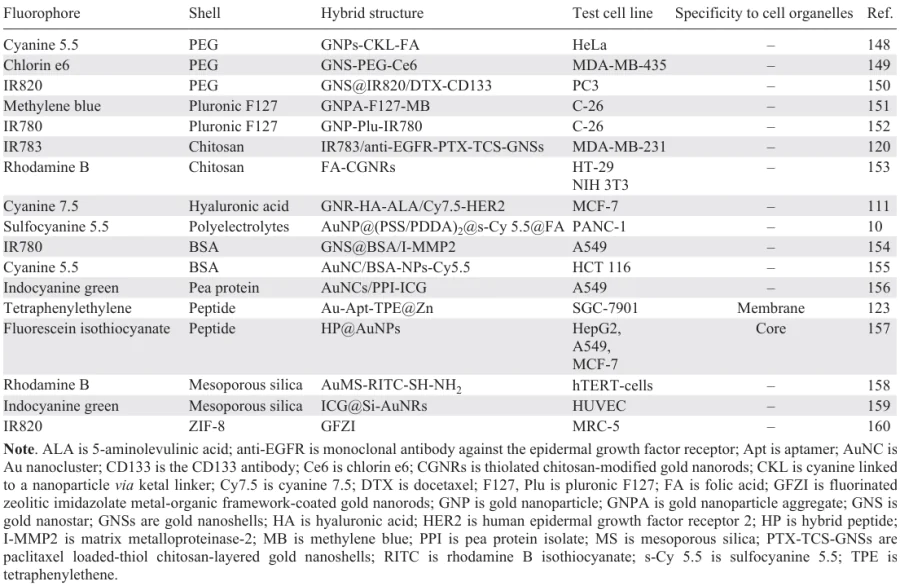
Selected examples of GNP-based hybrid structures developed for bioimaging are summarized in Table 3.[148-160] It can be seen that most of manufactured hybrid structures are non-specific to various cell organelles; in this case, the probes are located in cytosol. However, there are some examples where hybrid structures are endowed with specificity by attaching a vector peptide or protein, in particular affinity protein, to the cell membrane,[161-163] mitochondria,[164-166] lysosomes,[167-169] or the core.[157][170]
Also, it is necessary to mention yet few examples of in vitro tests of fluorescent gold-organic nanostructures using more complex cellular objects, spheroids,[171-177] which are simple models of biological systems. A publication by Sa´nchez et al.,[178] who studied fluorophore-labelled GNPs on U87MG glioblastoma cell spheroids, deserves attention. The authors were able to demonstrate that the hybrid structures based on polyaminocarboxylate-coated gold spheres of 2 – 3 nm in diameter conjugated with cyanine 5 N-hydroxysuccinimide derivative can reach the centre of a spheroid with a diameter of 800 μm (Fig. 10). This result is important for potential in vivo applications of hybrid nanostructures, as it shows their ability to be uniformly distributed throughout living tissues.
![[{"id":"EuSKLVqUEU","type":"paragraph","data":{"text":"(<i>a</i>) Fluorescence image of the U87 MG cell spheroid after 24-hour incubation with gold nanoparticles modified with diethylenetriaminepentaacetic acid bearing conjugated cyanine 5. The pink colour is due to nanoparticles localized in cytosol and the blue colour is caused by hoechst 33342 nucleus stain. (<i>b</i>) Fluorescence image of single U87 MG cells after dissociation of the spheroid.<sup>178</sup>"}}]](/storage/images/resized/fEVUW2Dn8qDVCYAqemXJtki0LOXZUksg7VKOYidn_xl.webp)
Fluorescence imaging using gold-organic nanoparticles can be performed not only in the confocal mode where fluorophore emission intensity is the measured parameter, but also in fluorescence lifetime imaging microscopy (FLIM) mode. FLIM has a higher specificity because the fluorophore lifetime is very sensitive to the environment, in particular, free fluorophore and fluorophore incorporated in a hybrid structure have different lifetimes. This fact is used to monitor the drug release if GNPs serve as carriers.[179] In addition, GNPs themselves can be imaged in this way in biological objects, which is of separate interest for cellular uptake studies.[180] For example, using particularly FLIM measurements, Han et al.[181] demonstrated that in small-sized PEG-coated GNPs functionalized with anti-EGFR antibodies, the spatial separation of the gold core and antibody coating takes place after endocytosis (Fig. 11). There are examples of bioimaging by muliphoton microscopy using GNP-and fluorophore-based hybrid nanostructures.[182][183]
![[{"id":"q_q9EPsDbq","type":"paragraph","data":{"text":"FLIM images of gold nanoparticles of 5 nm in diameter modified with anti-EGFR in A431 cells. (<i>a</i>) Fluorescence lifetime curves obtained in the cells from nanoparticles conjugated with unlabelled antibodies (green); cells incubated with AF647-labelled anti-EGFR-antibodies (red); and cells incubated with the cyan fluorescent protein (CFP) that was used to stain the plasmatic membrane of the cells (blue). The fluorescence lifetime in each case was measured without any other components. (<i>b</i>) FLIM image based on integral intensity. (<i>c</i>) FLIM image based on three lifetime components corresponding to the gold core (green), AF647-labelled antibodies (red), and cell membrane protein (blue). (<i>d</i>) FLIM images for each single component shown in (<i>c</i>). The incubation time of cells with nanoparticles was 24 h in all cases. The scale bars are 20 μm.<sup>181</sup> Figure reproduced with permission from the American Chemical Society."}}]](/storage/images/resized/yTLkRg4LLAAfq58Wp5mCHGnxRmIabFx8J4hIxHlD_xl.webp)
The dependence of the fluorophore lifetime on the distance to the gold core within the hybrid structure opens up the possibility of multiplexed analysis in FLIM.[48] Another way to achieve multiplexing by fluorescence microscopy (now in the confocal mode) is to fabricate hybrid probes with a set of vector–fluorophore combinations. An excellent example of these probes was reported by Lu et al.,[110] who conjugated spherical GNPs of 40 nm in diameter with three biomolecules (P) specific to various types of carcinogenic microRNA (mRNA) and functionalized with double-specific nucleases (DSN) and placed them into a cell membrane (CM) vesicle. Each vector biomolecule carried a different fluorescence probe with distinct working spectral ranges. In in vitro experiment, the authors clearly demonstrated the possibility of multiplexed determination of mRNA21, mRNA155, and mRNA205 in the MCF-7 cells using the prepared hybrid structures (Fig. 12).
![[{"id":"2TRdQX8SJU","type":"paragraph","data":{"text":"Fluorescence images of MCF-7 cells incubated with Au-P/DSN@CM and their precursors. Au-P/DSN@CM are gold nanoparticles functionalized with a vector (P) to definite mRNA and with double-specific nucleases and placed into a cell membrane (CM) vesicles. The signals of FAM (fluorescein), Cy3 (cyanine 3), and Cy5 (cyanine 5) fluorophores correspond to mRNA21 (green), mRNA155 (yellow), and mRNA205 (red). The signal appears when nanostructures bearing double-specific nucleases interact with mRNA and it is more intense for nanostructures occurring in CM vesicles, which provides higher cellular transfection efficiency. Down-regulation and up-regulation refer to decreasing or increasing mRNA expression attained by using small interfering RNA as regulators. The scale bar corresponds to 40 μm.<sup>110</sup> Figure reproduced with permission from the American Chemical Society. "}}]](/storage/images/resized/QZMTeLJy1yFlfFC0tz9jOhWVjjAp6L6WRdI53Mrn_xl.webp)
Apart from the agents for multiplexed bioimaging, agents with a multimodal response are particularly valuable for identifying the region of interest using a few methods. In the simplest cases, the multimodal response of a hybrid structure is provided by the properties of either fluorophore or the gold nanoparticles. In the former case, SERS method can also be used to select the optimal distance between the fluorophore and the gold particle.[184][185] In the latter case, owing to effective absorption of X-ray and optical radiation by GNPs, fluorescence imaging can be combined with computed tomography [186-191] and photoacoustic imaging.[192-195] The insertion of radioactive components into hybrid structures may give agents for fluorescence imaging coupled with positron emission tomography.[196-199] The use of gold composites with iron oxide, gadolinium or manganese compounds as the basis for fluorescence probes produces optically active contrasts suitable also for magnetic resonance imaging (MRI).[200-202] These compounds are the agents of choice as they are applicable in at least three independent bioimaging methods: fluorescence imaging, photoacoustic imaging, and MRI. In particular, Pan et al.[192] clearly demonstrated the benefits of complex agents of this type. Using a platform combining a composite based on gold nanocages and manganese dioxide with the cyanine 7 fluorophore, images of a mouse were obtained using each imaging technique; they demonstrate accumulation of the agent in the esophageal squamous cell carcinoma xenograft 24 h after intravenous administration (Fig. 13).
![[{"id":"5svGqwajuS","type":"paragraph","data":{"text":"(<i>a</i>) <i>In vivo</i> fluorescence images of a mouse before and after injection of the agent based on gold nanocages, manganese dioxide, and cyanine 7 (Cy7-FRNPs); (<i>b</i>) T1-weighted phantom images with different FRNP concentrations; (<i>c</i>) in vivo MRI images of the tumour site before and after the injection of FRNPs; (<i>d</i>) spin – lattice relaxation rates for solutions of FRNPs at different Mn<sup>2+</sup> concentrations; (<i>e</i>) <i>in vivo</i> photoacoustic image of the tumour site before and after injection of FRNPs.<sup>192</sup> Figure reproduced with permission from the Dove Medical Press."}}]](/storage/images/resized/dVdWCB0N3J6dfFyZn3TXfhSFQ00eViqfooqCmpNR_xl.webp)
5. Theranostics using hybrid gold-organic nanostructures
The GNP- and fluorophore-based structures can be used to design theranostic platforms, which, apart from the bioimaging diagnostic function, possess a therapeutic effect. Platforms of this type are designed for photothermal therapy, photodynamic therapy, and chemotherapy; PTT is considered to be the main application of GNP-based agents.
An advantage of PTT is that it can be performed locally. Therefore, GNPs can be considered to be promising for the treatment of malignant tumours of the brain and central nervous system where it is very important to minimize the effect on healthy tissues, which is not the case for conventional surgery techniques, chemotherapy, and radiotherapy. The high potential of GNPs for neuro-oncology has already brought the relevant in vivo studies to a rather high level and would stimulate the development of related technical equipment to perform local PTT. In particular, Arami et al.[203] recently reported a method for remotely controlled PTT of a brain tumour for freely behaving mice using PEG-stabilized gold nanostars equipped with a fluorescence and Raman probes for imaging.[203] A specific feature of the presented procedure is that local irradiation of gold nanostars after their intratumoral injection is performed using a miniature subcutaneously implanted flexible near-IR light-emitting device, which is controlled using near field communication (NFC) technology. This device made it possible to carry out periodic PTT without anesthesia for 15 days; a considerable decrease in the tumour size was detected by histological analysis. The authors also demonstrated local heating of only the tumour area in the temperature images of the open skull of an individual animal.
For about a decade, PTT with GNP-based agents has been conducted using, in particular, large laboratory animals. An early study [204] reports the results of therapy of 13 mammary tumours in Persian cats and griffon dogs with PEG-stabilized gold nanorods with an aspect ratio of approximately five. As shown by the authors, irradiation for two minutes repeated three times at two-week intervals with a laser power density of 5.8 mW cm–2 is sufficient for complete regression of the tumour (Fig. 14). An important supplement of the study is experimental demonstration of increasing proportion of cell death by necrosis rather than apoptosis with increasing irradiation time.
![[{"id":"ags5d6y9oA","type":"paragraph","data":{"text":"Photographs of a dog and histopathological images demonstrating the tumour state before (<i>a</i>) and after (<i>b</i>) the therapy.<sup>204</sup> Figure reproduced with permission from Dove Medical Press."}}]](/storage/images/resized/FouA39uy4ba9yWaz22bzTBLakSo4hPQS4ZNfaaVJ_xl.webp)
Schuh et al.[205] also performed PTT using PEG-stabilized gold nanorods for dogs differing in the breed, age, sex, and weight with spontaneous malignant tumours. The study demonstrated low toxicity of the medication for all animals, pronounced photothermal effect sufficient for conducting the therapy of even relatively large malignant tumours. The adverse events were restricted to local reactions to laser irradiation at 808 nm. Important observations are concerned with the circulation of gold nanorods in the body, which showed that they can be completely eliminated from the bloodstream after 72 h. Similar results were obtained in later studies dealing with the action of gold nanorods on laboratory mice.[206-208]
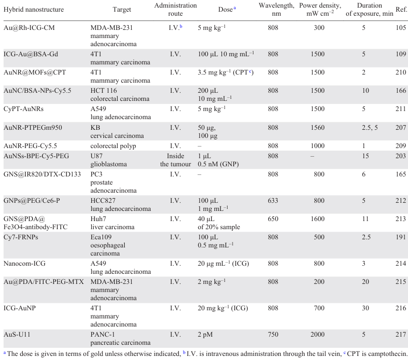
Studies describing the use of GNP- and fluorophore-based medications for the therapy of large laboratory animals are still rather scarce; however, experiments on mice and rats are carried out throughout the world. Almost each case demonstrates the high photothermal efficiency and benefits of incorporation of fluorophores for the imaging of introduced GNPs. In particular, Gournaris et al.[209] used an endoscopic fluorescence imaging system for real-time monitoring of the gradual change in the size of the colorectal polyp during GNP-mediated thermal ablation. Selected in vivo studies using GNP- and fluorophore-based systems are given in Table 4;[210-217] they reveal some issues that have to be discussed. First, GNP-based hybrid structures can be targeted to tumours of different types and locations; most often, it is assumed that the agents are administered intravenously or injected directly into the tumour. Second, there is a rather strong variation in the doses of gold used. Furthermore, there is actually no uniformity in the indication of the doses. In the case of intravenous administration, the dose should be related to the weight of the laboratory animal; however, the authors often neglect this and indicate only the volume and concentration of the injected dispersion. Moreover, in some cases, the weight concentration of gold is indicated, while in other cases, the numerical concentration of particles is given. When GNPs are injected directly into the tumour, comparison of the doses used in different studies is even less obvious. In this case, the dose must be correlated with the tumour volume, which is often unknown. Therefore, currently, we can only make a general conclusion that for intravenous administration of gold-containing drugs for PTT, the dose of the drug related to the weight of the laboratory animal often falls within the range of 2 – 5 mg kg–1.
The hybrid structure consisting of GNPs and indocyanine green (see Table 4) [109] is a vivid example of a theranostic agent not only for the combined imaging and therapy, but also for the combined photothermal and photodynamic therapy. A number of studies demonstrate that GNPs can enhance generation of reactive oxygen species (ROS) in some molecules.[218][219] Owing to this feature, active development of GNP-based hybrid structures containing also photosensitizers is currently in progress.[220][221] Gold nanorods bound through a linker (thioglycolic acid) to the mitochondrially targeted heptamethinecyanine fluorophore is an example of such structure.[211] These structures make it possible to observe the heptamethinecyanine fluorescence in vivo; they possess photothermal and photodynamic effects and have high sensitivity to glutathione and, hence, they can be used to determine the glutathione concentration. Another bright example of hybrid nanostructures with integrated therapeutic effect and the possibility of fluorescence bioimaging is the theranostic platform based on gold nanorods, metal-organic framework of zirconium clusters, and carboxyl tetraphenylporphyrin derivative loaded with a topoisomerase inhibitor, camptothecin.[210] An in vivo test of this agent showed high efficacy against mammary carcinoma confirmed by analysis of the survival rate of mice and the weight of tumours excised after the therapy.
The photothermal effect of GNPs is used not only for local hyperthermia of cancer cells, but also for photocontrolled drug release if the hybrid structure contains a temperature-sensitive polymer. The Nanocom-ICG drug (authors’ designation) [214] consisting of gold nanorods with a poly(N-isopropylacrylamide) shell containing ICG is a demonstration of this approach. According to experimental data of the authors, the photothermal heating of nanostructures is accompanied by degradation of the shell, resulting in the release of indocyanine green and aggregation of GNPs. Finally, indocyanine green molecules penetrate directly into cancer cells, thus generating ROS inside them, while the photothermal effect and photoacoustic signal of GNPs are enhanced (Fig. 15).
![[{"id":"y4rIVQm0wh","type":"paragraph","data":{"text":"(<i>a</i>) Infrared images of A549 tumour-bearing mice after laser irradiation at 808 nm (0.8 W cm<sup>–2</sup>, 3 min) three days after the administration of Nanocom-ICG or its components. (<i>b</i>) Change in the tumour volume and (<i>c</i>) survival rate of mice in various groups after the therapy (number of repetitions n = 5, *p < 0.5, **p < 0.05, p-confidence levels: 0.5 is the border of statistical significance, 0.05 corresponds to a statistically significant difference). (<i>d</i>) Photographs of the tumours taken from animals of different groups 21 day after the therapy. (<i>e</i>) Western blotting assay of the expression of HSP90 and HSP70 heat shock proteins in tumour tissues after irradiation.<sup>214</sup> Figure reproduced with permission from Springer Nature."}}]](/storage/images/resized/ntxku067xmW0LPIR9tjQlapnzLs2VwgXnWX1wTWU_xl.webp)
Gold nanovesicles are of interest as integrated theranostic platforms in which PTT is supplemented by chemotherapy. Their benefits include not only excellent photothermal properties, but also high drug loading capacity.[222-225] Deng et al.[226] reported gold nanovesicles loaded with a chemotherapy drug, which were fabricated by the assembly of PEG – polycaprolactone block copolymer-coated GNPs and encapsulation of doxorubicin. In in vivo experiments on mice, the use of these nanovesicles resulted in complete elimination of glioblastoma.
An important issue in the development of gold-containing theranostic drugs is their targeting of specific biological sites, which can considerably reduce the overall burden on the body. For the targeted delivery, GNPs are most often functionalized with vector molecules such as proteins, low-molecular-weight polypeptides, and growth factors. A wide variety of protein molecules can be used, for exampple, antibodies such as immunoglobulins G (IgG).[227][228] Among low-molecular-weight peptides, mention should be made of argynylglycylaspartic acid (RGD sequence) tripeptide derivatives used to deliver GNPs to cell lines characterized by overexpression of the membrane proteins integrins (e.g., lung cancer or mammary cancer cells).[229] Small peptide molecules known as affibodies also deserve attention.[230] They are analogues of monoclonal antibodies with similar specificity to cancer cells and smaller size. This fact facilitates nanoparticle modification by these species and increases the colloidal stability of the resulting structures.[231][232] Growth factors, which are essential for cell development and are consumed in increased amounts by some cancer cells, are also being investigated as vectors for the delivery of nanoplatforms; among them, folic acid is widely used.[233][234]
There is a special approach to the delivery of GNPs (and any other drug) known as the bioorthogonal chemistry.[235] This method implies in situ conduction of a highly specific and biocompatible reaction to conjugate particles to the affected cells.[236] The list of functional groups used in bioorthogonal chemistry largely overlaps with those discussed in Section 3.2 in relation to covalent conjugation of fluorophores to GNPs; however, in this case, the reactions of azides with alkynes and reactions of tetrazines with dienophiles are utilized most often.[237][238]
Unfortunately, the above examples of successful animal trials of GNP-based drugs and some success in the development of their targeted delivery do not eliminate a number of problems that preclude switching to their larger-scale use in vivo. The major obstacle to the clinical use of GNPs (as well as other inorganic nanoparticles) is the risk of thrombosis and delayed side effects. Numerous independent clinical trials are needed to answer the questions related to the biosafety of GNPs. A review by Yao et al.[239] summarizes the results of more than 20 studies devoted to the use of GNPs as theranostic agents. The authors concluded that GNPs are safe for humans, but at the same time, they pointed out the limited amount of data: the small number of trials including small numbers of subjects.
Special mention should be made of the limitations and disadvantages of photothermal and photodynamic therapy, which are direct methods for implementation of the therapeutic potential of GNPs. The key limitation of PTT and PDT, caused by the small depth of penetration of optical and NIR radiation into tissues, is that only surface or near-surface structures can be treated. Another common problem of the two methods is the lack of safety of using long-wavelength lasers, which is a challenge both for patients and for doctors. In the case of PTT, a complicated issue is to accurately measure the local temperature upon laser treatment in order to avoid the tissue overheating and, as a consequence, necrosis. Another drawback of PTT is the painfulness of the procedure, with the use of anesthesia being impossible. The limitations of PTT, in particular with the use of GNPs, were considered in detail by Salimi et al.[240] One of the most severe problems of PDT is the possible tissue hypoxia, which markedly decreases the efficiency of the procedure.
6. Conclusion
Combining gold nanoparticles and molecular fluorophores into hybrid structures is an attractive strategy for the design of nano-sized agents for biomedical purposes. However, due to the complex influence of plasmon resonance on the photophysical properties of fluorophores, this approach requires thorough optimization of the hybrid structures, in particular the fluorophore position relative to the metal core. Currently, methods of fabrication of hybrid structures using molecular linkers or polymeric or inorganic coatings as well as DNA origami technique are successfully coping with this task.
The effective GNP-based hybrid systems are being developed in an integrated manner, using anisotropic particles or their small aggregates and coatings that promote targeted delivery, increase the payload, and decrease the toxicity. However, regarding the optical issues, the design of bioimaging and therapy agents based on GNPs and fluorophores is delayed by the small number of fluorophores for the NIR range. Currently, structures containing indocyanine green are investigated almost without alternatives in this range. Despite the examples of hybrid structures with other NIR fluorophores reported in the literature, there is problem of exceptionally low photostability of these compounds.
A key motivation for the research of hybrid structures based on GNPs and fluorophores is their obvious advantage of multimodality and possibility of providing an integrated effect, which is much more difficult to achieve in the case of molecular agents. This is responsible for the general trend that can be followed in recent studies, that is, design of complex theranostic platforms combining several bioimaging and therapeutic agents. However, it should be borne in mind that as the functions of such platforms are expanded, the reproducible production of the platforms becomes more complicated and the problem of low colloidal stability becomes more acute.
The safety of using GNPs and hybrid structures based on them in living organisms remains an open question. Currently, even studies on cell lines do not provide unambiguous information on what is the threshold of acceptable content of gold nanoparticles before they show toxic effects. The reported values of the weight concentrations of gold or numerical concentration of nanoparticles differ significantly between various publications. Nevertheless, quite a few successful examples of in vitro studies on cell lines and in vivo studies on mice have been reported in the literature, demonstrating effective optical diagnosis and photothermal therapy using gold-organic nanostructures. However, currently, there are still few worthy examples of in vivo tests implemented on large laboratory animals. Certainly, this situation may be associated, to some extent, with high cost of these studies, but this mere fact raises some doubts about near future implementation of GNP- and fluorophore-based agents into practical medicine. In this regard, the few publications by leading research groups that were carried out with dogs and that can be considered as predecessors for further clinical trials are encouraging.
On the whole, theranostics with hybrid structures based on GNPs and fluorophores is certainly highly attractive for medicine, but there are still a lot challenges to be overcomed before they can be implemented in practice. In addition, major advances are needed in the technical equipment for illumination and signal recording, such as miniature endoscopic imaging systems or implantable local illumination devices. The studies discussed in this review indicate that necessary efforts are being made by scientific community in cooperation with technological companies and healthcare facilities, which ensures the slow progress in the implementation of nanostructures based on GNPs and fluorophores in practical medicine.
This review was prepared with the financial support of the Saint Petersburg State University (Project No. 122040800256-8, Sections 1, 2, 5 – 6) and the Russian Science Foundation (Project No. 22-73-10052, Sections 3, 4).
7. List of abbreviations and symbols
AIE — aggregation-induced emission,
BI — bioimaging,
BDP — boron dipyrromethene,
BSA — bovine serum albumin,
CM — cell membrane,
DSN — double-specific nucleases,
FAM — fluorescein,
FLIM — fluorescence lifetime imaging microscopy,
FRET — Förster resonance energy transfer,
GNPs — gold nanoparticles,
ICG — indocyanine green,
LSPR — localized surface plasmon resonance,
mRNA — microRNA,
MRI — magnetic resonance imaging,
MHR — molar heating rate,
NIR — near infrared range,
NSET — nanosurface energy transfer,
PDT — photodynamic therapy,
PDDA — poly(diallyldimethylammonium chloride),
PEG — polyethylene glycol,
PSS — poly(sodium styrenesulfonate),
PTT — photothermal therapy,
ROS — reactive oxygen species,
SERS — surface-enhanced Raman scattering,
SPCE — surface plasmon-coupled emission,
a — surface curvature radius,
A — rate of light energy absorption by the irradiated system,
Aλ — absorbance of a nanoparticle at the excitation wavelength,
B — energy dissipation rate,
Ci — heat capacity of the ith component,
D — diameter of a metal sphere,
d — distance to the surface,
E — vector of the local electric field near the plasmonic particle,
E0 — incident electric field vector,
Eabs — heating caused by absorption by particles,
Eloss — heat lost to the environment,
F — fluorescence intensity of a free molecule,
F0 — fluorescence intensity of a molecule in the presence of a metallic nanostructure,
f — directionality factor of fluorophore emission,
h — heat transfer coefficient,
kD — rate constant for the nanoparticle–fluorophore energy transfer,
ke — rate constant for the fluorophore excitation in the electric field of the nanoparticle,
ke0 — rate constant for excitation of free fluorophore in a homogeneous medium,
λabs — absorption wavelength,
λem — emission wavelength,
λex — excitation wavelength,
kabs — constant for the non-radiative decay of the fluorophore excited state in the nanoparticle electric field via thermal dissipation of energy,
km — constant for the non-radiative decay of the fluorophore excited state in the nanoparticle electric field via binding to non-radiative electromagnetic modes,
kr — constant for the radiative decay of the fluorophore excited state in the nanoparticle electric field,
kr0 — constant for the radiative decay of the free fluorophore excited state in a homogeneous medium,
knr —constant for the non-radiative decay of the fluorophore excited state in the nanoparticle electric field,
knr0 — constant for the non-radiative decay of the free fluorophore excited state in a uniform field,
MEM — electromagnetic fluorescence enhancement factor,
mi — weight of the ith component,
R — distance from the point to the centre of the metal sphere,
R0 — distance at which the efficiency of energy transfer is 50%,
P — laser power,
τD — fluorescence lifetime in the presence of an acceptor (nanoparticle),
S — surface area covered by the nanoparticle dispersion,
T0 — ambient temperature,
Φ0 — fluorescence quantum yield,
Φ — fluorescence quantum yield near the plasmonic surface,
μabs — dipole moment of the transition corresponding to radiation absorption,
μem — dipole moment of the transition corresponding to emission,
η — photothermal conversion efficiency.
References